| << Chapter < Page | Chapter >> Page > |
Fluorescence can be induced by many types of energy input. Fluorescent paint, dyes, and even soap residues in clothes make colors seem brighter in sunlight by converting some UV into visible light. X rays can induce fluorescence, as is done in x-ray fluoroscopy to make brighter visible images. Electric discharges can induce fluorescence, as in so-called neon lights and in gas-discharge tubes that produce atomic and molecular spectra. Common fluorescent lights use an electric discharge in mercury vapor to cause atomic emissions from mercury atoms. The inside of a fluorescent light is coated with a fluorescent material that emits visible light over a broad spectrum of wavelengths. By choosing an appropriate coating, fluorescent lights can be made more like sunlight or like the reddish glow of candlelight, depending on needs. Fluorescent lights are more efficient in converting electrical energy into visible light than incandescent filaments (about four times as efficient), the blackbody radiation of which is primarily in the infrared due to temperature limitations.
This atom is excited to one of its higher levels by absorbing a UV photon. It can de-excite in a single step, re-emitting a photon of the same energy, or in several steps. The process is called fluorescence if the atom de-excites in smaller steps, emitting energy different from that which excited it. Fluorescence can be induced by a variety of energy inputs, such as UV, x-rays, and electrical discharge.
The spectacular Waitomo caves on North Island in New Zealand provide a natural habitat for glow-worms. The glow-worms hang up to 70 silk threads of about 30 or 40 cm each to trap prey that fly towards them in the dark. The fluorescence process is very efficient, with nearly 100% of the energy input turning into light. (In comparison, fluorescent lights are about 20% efficient.)
Fluorescence has many uses in biology and medicine. It is commonly used to label and follow a molecule within a cell. Such tagging allows one to study the structure of DNA and proteins. Fluorescent dyes and antibodies are usually used to tag the molecules, which are then illuminated with UV light and their emission of visible light is observed. Since the fluorescence of each element is characteristic, identification of elements within a sample can be done this way.
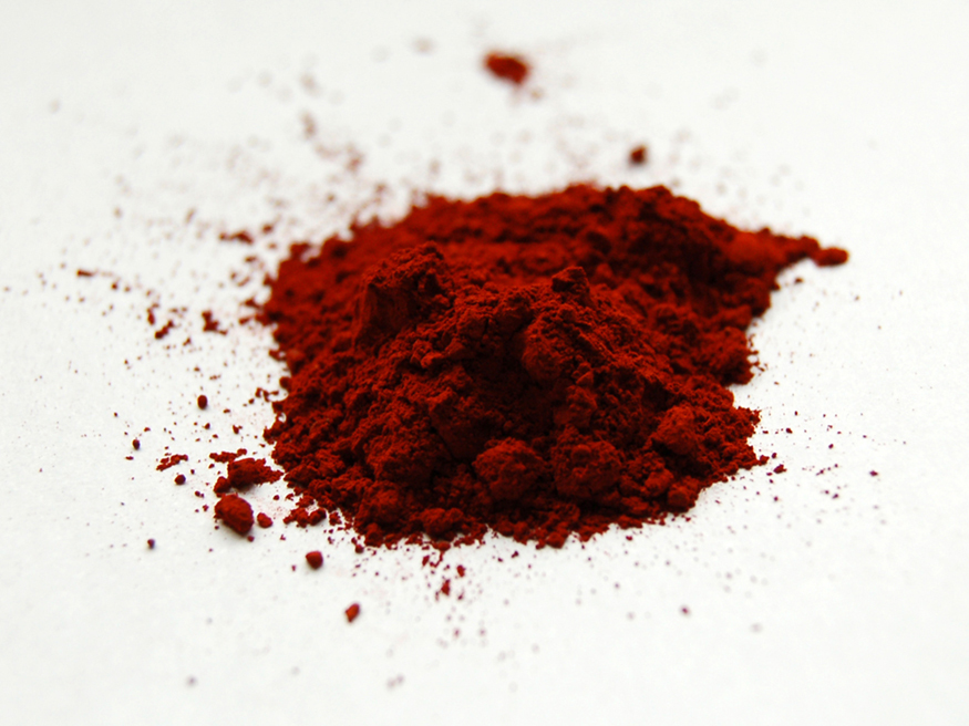
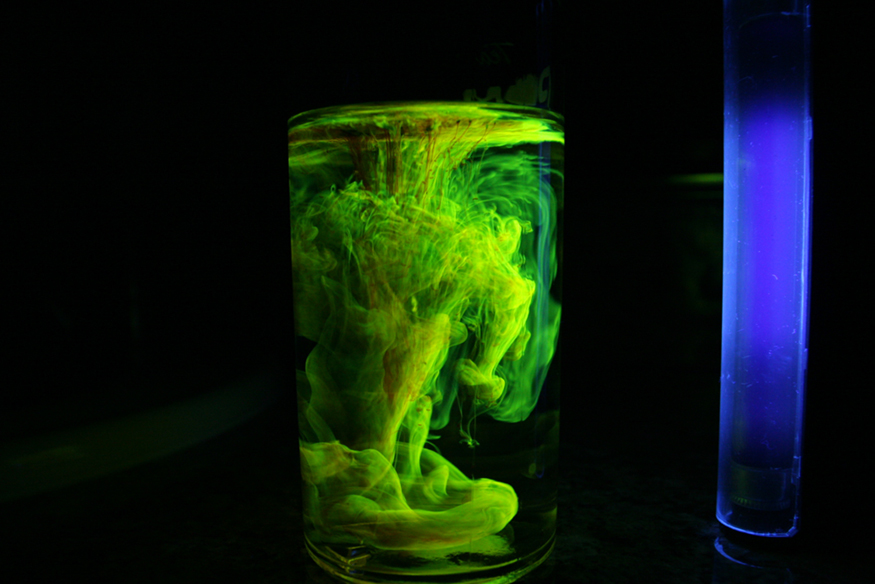
[link] shows a commonly used fluorescent dye called fluorescein. Below that, [link] reveals the diffusion of a fluorescent dye in water by observing it under UV light.


Recently, a new class of fluorescent materials has appeared—“nano-crystals.” These are single-crystal molecules less than 100 nm in size. The smallest of these are called “quantum dots.” These semiconductor indicators are very small (2–6 nm) and provide improved brightness. They also have the advantage that all colors can be excited with the same incident wavelength. They are brighter and more stable than organic dyes and have a longer lifetime than conventional phosphors. They have become an excellent tool for long-term studies of cells, including migration and morphology. ( [link] .)

Notification Switch
Would you like to follow the 'Basic physics for medical imaging' conversation and receive update notifications?