<< Chapter < Page Chapter >> Page >

The two suture lines seen on the top of the skull are the coronal and sagittal sutures. The coronal suture    runs from side to side across the skull, within the coronal plane of section (see [link] ). It joins the frontal bone to the right and left parietal bones. The sagittal suture    extends posteriorly from the coronal suture, running along the midline at the top of the skull in the sagittal plane of section (see [link] ). It unites the right and left parietal bones. On the posterior skull, the sagittal suture terminates by joining the lambdoid suture. The lambdoid suture    extends downward and laterally to either side away from its junction with the sagittal suture. The lambdoid suture joins the occipital bone to the right and left parietal and temporal bones.

Facial bones of the skull

The facial bones of the skull form the upper and lower jaws, the nose, nasal cavity and nasal septum, and the orbit. The facial bones include 14 bones, with six paired bones and two unpaired bones. The paired bones are the maxilla, palatine, zygomatic, nasal, lacrimal, and inferior nasal conchae bones. The unpaired bones are the vomer and mandible bones. Although classified with the brain-case bones, the ethmoid bone also contributes to the nasal septum and the walls of the nasal cavity and orbit.

Maxillary bone

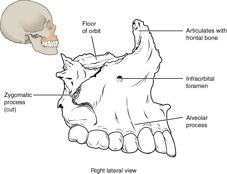
The maxillary bone    , often referred to simply as the maxilla (plural = maxillae), is one of a pair that together form the upper jaw, much of the hard palate, and the base of the nose (see [link] ). Each tooth is anchored into a deep socket called an alveolus. . The hard palate    is the bony plate that forms the roof of the mouth and floor of the nasal cavity, separating the oral and nasal cavities.
Maxillary bone

This image shows the location and structure of the maxilla. A small image of the skull on the top left shows the maxilla in ochre yellow. A magnified view shows the detailed structure of the maxilla.
The maxillary bone forms the upper jaw and supports the upper teeth. Each maxilla also forms the lateral floor of each orbit and the majority of the hard palate.

Zygomatic bone

The zygomatic bone    is also known as the cheekbone. Each of the paired zygomatic bones forms much of the wall of the orbit.

Nasal bone

The nasal bone    is one of two small bones that articulate (join) with each other to form the bony base (bridge) of the nose. They also support the cartilages that form the lateral walls of the nose (see [link] ). These are the bones that are damaged when the nose is broken.

Lacrimal bone

Each lacrimal bone    is a small, rectangular bone that forms the anterior, medial wall of the orbit This bone contains the nasalacrimal canal, through which tears pass.

Vomer bone

The unpaired vomer bone, often referred to simply as the vomer, is triangular-shaped and forms the lower part of the nasal septum (see [link] ).

Mandible

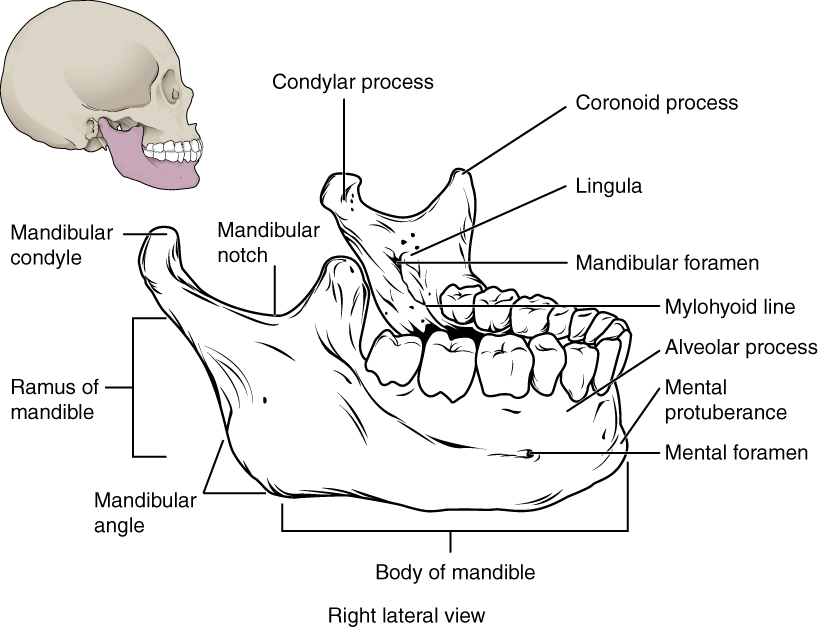
The mandible    forms the lower jaw and is the only moveable bone of the skull. At the time of birth, the mandible consists of paired right and left bones, but these fuse together during the first year to form the single U-shaped mandible of the adult skull.
Isolated mandible

This image shows the structure of the mandible. On the top left, a lateral view of the skull shows the location of the mandible in purple. A magnified image shows the right lateral view of the mandible with the major parts labeled.
The mandible is the only moveable bone of the skull.

Paranasal sinuses

The paranasal sinuses    are hollow, air-filled spaces located within certain bones of the skull ( [link] ). All of the sinuses communicate with the nasal cavity (paranasal = “next to nasal cavity”) and are lined with nasal mucosa. They serve to reduce bone mass and thus lighten the skull, and they also add resonance to the voice. This second feature is most obvious when you have a cold or sinus congestion. These produce swelling of the mucosa and excess mucus production, which can obstruct the narrow passageways between the sinuses and the nasal cavity, causing your voice to sound different to yourself and others. This blockage can also allow the sinuses to fill with fluid, with the resulting pressure producing pain and discomfort.

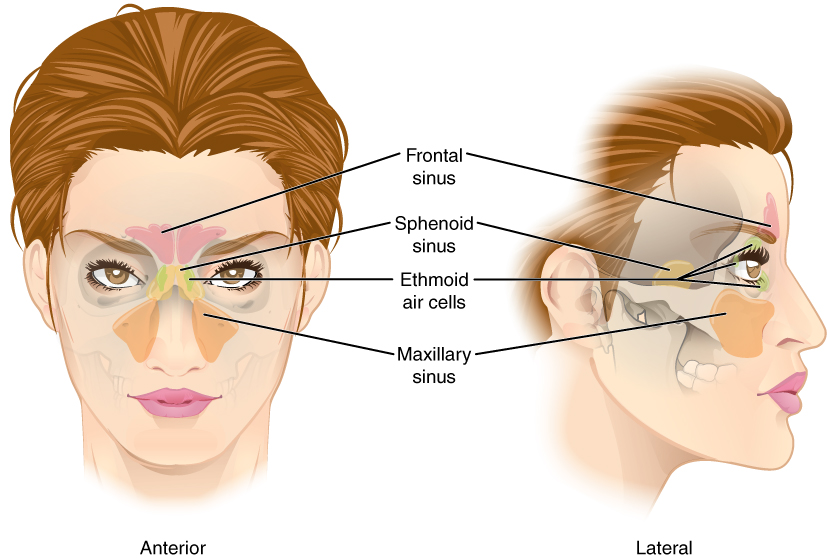
The paranasal sinuses are named for the skull bone that each occupies. The frontal sinus    is located just above the eyebrows, within the frontal bone (see [link] ). The largest sinus is the maxillary sinus    . These are paired and located within the right and left maxillary bones, where they occupy the area just below the orbits. The maxillary sinuses are most commonly involved during sinus infection because of their close connection to the nasal cavity. The sphenoid sinus    is a single, midline sinus. The ethmoid bone contains multiple small spaces separated by very thin bony walls. These are collectively called the ethmoid sinus .
Paranasal sinuses

This figure shows a woman’s face and the location of the paranasal sinuses. The left panel shows the anterior view of the woman’s face with the sinuses labeled. The right panel shows the lateral view of the woman’s face with the same parts labeled.
The paranasal sinuses are hollow, air-filled spaces named for the skull bone that each occupies. The most anterior is the frontal sinus, located in the frontal bone above the eyebrows. The largest are the maxillary sinuses, located in the right and left maxillary bones below the orbits. The most posterior is the sphenoid sinus, located in the body of the sphenoid bone, under the sella turcica. The ethmoid air cells are multiple small spaces located in the right and left sides of the ethmoid bone, between the medial wall of the orbit and lateral wall of the upper nasal cavity.

Hyoid bone

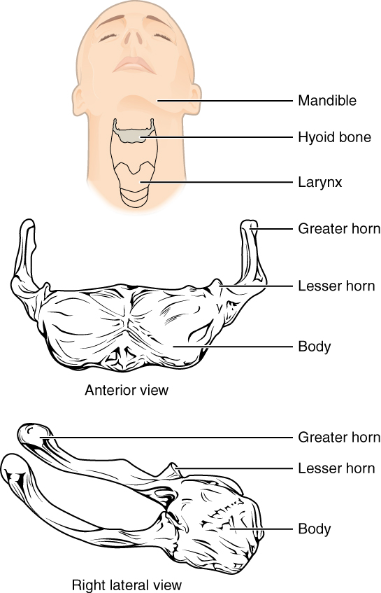
The hyoid bone is an independent bone that does not contact any other bone and thus is not part of the skull ( [link] ). It is a small U-shaped bone located in the upper neck near the mandible, with the tips of the “U” pointing backwards. The hyoid serves as the base for the tongue above, and is attached to the larynx and the pharynx .
Hyoid bone

In this image, the location and structure of the hyoid bone are shown. The top panel shows a person’s face and neck, with the hyoid bone highlighted in grey. The middle panel shows the anterior view and the bottom panel shows the right anterior view.
The hyoid bone is located in the upper neck and does not join with any other bone. It provides attachments for muscles that act on the tongue, larynx, and pharynx.

References

Centers for Disease Control and Prevention (US). Injury prevention and control: traumatic brain injury [Internet]. Atlanta, GA; [cited 2013 Mar 18]. Available from: (External Link) .

Questions & Answers

what is microbiology
Agebe Reply
What is a cell
Odelana Reply
what is cell
Mohammed
how does Neisseria cause meningitis
Nyibol Reply
what is microbiologist
Muhammad Reply
what is errata
Muhammad
is the branch of biology that deals with the study of microorganisms.
Ntefuni Reply
What is microbiology
Mercy Reply
studies of microbes
Louisiaste
when we takee the specimen which lumbar,spin,
Ziyad Reply
How bacteria create energy to survive?
Muhamad Reply
Bacteria doesn't produce energy they are dependent upon their substrate in case of lack of nutrients they are able to make spores which helps them to sustain in harsh environments
_Adnan
But not all bacteria make spores, l mean Eukaryotic cells have Mitochondria which acts as powerhouse for them, since bacteria don't have it, what is the substitution for it?
Muhamad
they make spores
Louisiaste
what is sporadic nd endemic, epidemic
Aminu Reply
the significance of food webs for disease transmission
Abreham
food webs brings about an infection as an individual depends on number of diseased foods or carriers dully.
Mark
explain assimilatory nitrate reduction
Esinniobiwa Reply
Assimilatory nitrate reduction is a process that occurs in some microorganisms, such as bacteria and archaea, in which nitrate (NO3-) is reduced to nitrite (NO2-), and then further reduced to ammonia (NH3).
Elkana
This process is called assimilatory nitrate reduction because the nitrogen that is produced is incorporated in the cells of microorganisms where it can be used in the synthesis of amino acids and other nitrogen products
Elkana
Examples of thermophilic organisms
Shu Reply
Give Examples of thermophilic organisms
Shu
advantages of normal Flora to the host
Micheal Reply
Prevent foreign microbes to the host
Abubakar
they provide healthier benefits to their hosts
ayesha
They are friends to host only when Host immune system is strong and become enemies when the host immune system is weakened . very bad relationship!
Mark
what is cell
faisal Reply
cell is the smallest unit of life
Fauziya
cell is the smallest unit of life
Akanni
ok
Innocent
cell is the structural and functional unit of life
Hasan
is the fundamental units of Life
Musa
what are emergency diseases
Micheal Reply
There are nothing like emergency disease but there are some common medical emergency which can occur simultaneously like Bleeding,heart attack,Breathing difficulties,severe pain heart stock.Hope you will get my point .Have a nice day ❣️
_Adnan
define infection ,prevention and control
Innocent
I think infection prevention and control is the avoidance of all things we do that gives out break of infections and promotion of health practices that promote life
Lubega
Heyy Lubega hussein where are u from?
_Adnan
en français
Adama
which site have a normal flora
ESTHER Reply
Many sites of the body have it Skin Nasal cavity Oral cavity Gastro intestinal tract
Safaa
skin
Asiina
skin,Oral,Nasal,GIt
Sadik
How can Commensal can Bacteria change into pathogen?
Sadik
How can Commensal Bacteria change into pathogen?
Sadik
all
Tesfaye
by fussion
Asiina
what are the advantages of normal Flora to the host
Micheal
what are the ways of control and prevention of nosocomial infection in the hospital
Micheal
Got questions? Join the online conversation and get instant answers!
Jobilize.com Reply

Get Jobilize Job Search Mobile App in your pocket Now!

Get it on Google Play Download on the App Store Now


Source:  OpenStax, Skeletal system. OpenStax CNX. Apr 17, 2015 Download for free at https://legacy.cnx.org/content/col11779/1.1
Google Play and the Google Play logo are trademarks of Google Inc.

Notification Switch

Would you like to follow the 'Skeletal system' conversation and receive update notifications?

Ask