<< Chapter < Page Chapter >> Page >

The two suture lines seen on the top of the skull are the coronal and sagittal sutures. The coronal suture    runs from side to side across the skull, within the coronal plane of section (see [link] ). It joins the frontal bone to the right and left parietal bones. The sagittal suture    extends posteriorly from the coronal suture, running along the midline at the top of the skull in the sagittal plane of section (see [link] ). It unites the right and left parietal bones. On the posterior skull, the sagittal suture terminates by joining the lambdoid suture. The lambdoid suture    extends downward and laterally to either side away from its junction with the sagittal suture. The lambdoid suture joins the occipital bone to the right and left parietal and temporal bones.

Facial bones of the skull

The facial bones of the skull form the upper and lower jaws, the nose, nasal cavity and nasal septum, and the orbit. The facial bones include 14 bones, with six paired bones and two unpaired bones. The paired bones are the maxilla, palatine, zygomatic, nasal, lacrimal, and inferior nasal conchae bones. The unpaired bones are the vomer and mandible bones. Although classified with the brain-case bones, the ethmoid bone also contributes to the nasal septum and the walls of the nasal cavity and orbit.

Maxillary bone

The maxillary bone    , often referred to simply as the maxilla (plural = maxillae), is one of a pair that together form the upper jaw, much of the hard palate, and the base of the nose (see [link] ). Each tooth is anchored into a deep socket called an alveolus. . The hard palate    is the bony plate that forms the roof of the mouth and floor of the nasal cavity, separating the oral and nasal cavities.
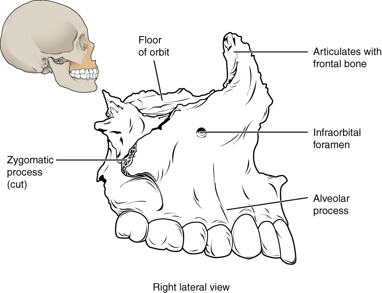
Maxillary bone

This image shows the location and structure of the maxilla. A small image of the skull on the top left shows the maxilla in ochre yellow. A magnified view shows the detailed structure of the maxilla.
The maxillary bone forms the upper jaw and supports the upper teeth. Each maxilla also forms the lateral floor of each orbit and the majority of the hard palate.

Zygomatic bone

The zygomatic bone    is also known as the cheekbone. Each of the paired zygomatic bones forms much of the wall of the orbit.

Nasal bone

The nasal bone    is one of two small bones that articulate (join) with each other to form the bony base (bridge) of the nose. They also support the cartilages that form the lateral walls of the nose (see [link] ). These are the bones that are damaged when the nose is broken.

Lacrimal bone

Each lacrimal bone    is a small, rectangular bone that forms the anterior, medial wall of the orbit This bone contains the nasalacrimal canal, through which tears pass.

Vomer bone

The unpaired vomer bone, often referred to simply as the vomer, is triangular-shaped and forms the lower part of the nasal septum (see [link] ).

Mandible

The mandible    forms the lower jaw and is the only moveable bone of the skull. At the time of birth, the mandible consists of paired right and left bones, but these fuse together during the first year to form the single U-shaped mandible of the adult skull.
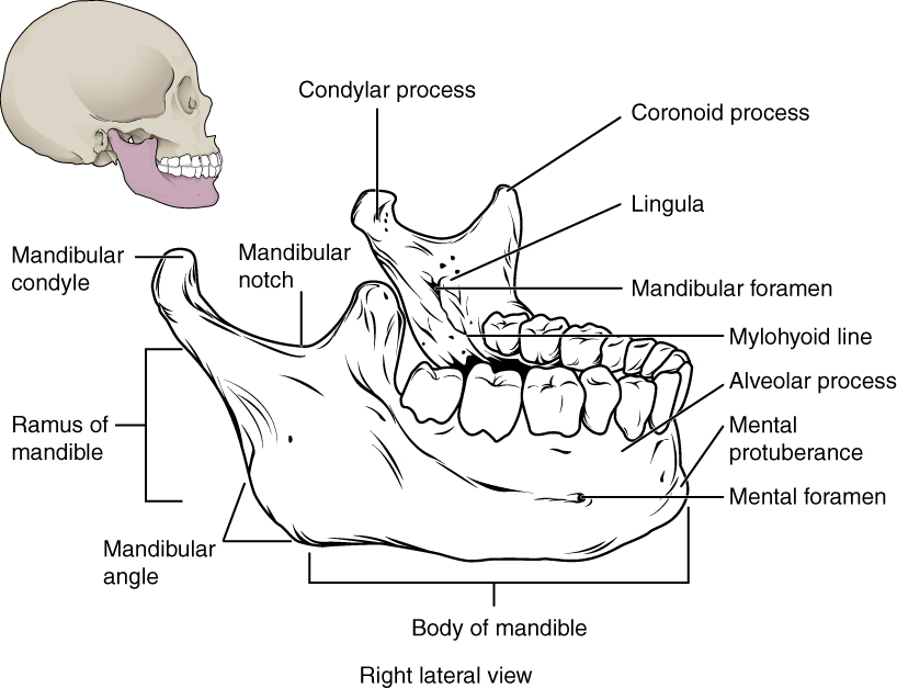
Isolated mandible

This image shows the structure of the mandible. On the top left, a lateral view of the skull shows the location of the mandible in purple. A magnified image shows the right lateral view of the mandible with the major parts labeled.
The mandible is the only moveable bone of the skull.

Paranasal sinuses

The paranasal sinuses    are hollow, air-filled spaces located within certain bones of the skull ( [link] ). All of the sinuses communicate with the nasal cavity (paranasal = “next to nasal cavity”) and are lined with nasal mucosa. They serve to reduce bone mass and thus lighten the skull, and they also add resonance to the voice. This second feature is most obvious when you have a cold or sinus congestion. These produce swelling of the mucosa and excess mucus production, which can obstruct the narrow passageways between the sinuses and the nasal cavity, causing your voice to sound different to yourself and others. This blockage can also allow the sinuses to fill with fluid, with the resulting pressure producing pain and discomfort.

The paranasal sinuses are named for the skull bone that each occupies. The frontal sinus    is located just above the eyebrows, within the frontal bone (see [link] ). The largest sinus is the maxillary sinus    . These are paired and located within the right and left maxillary bones, where they occupy the area just below the orbits. The maxillary sinuses are most commonly involved during sinus infection because of their close connection to the nasal cavity. The sphenoid sinus    is a single, midline sinus. The ethmoid bone contains multiple small spaces separated by very thin bony walls. These are collectively called the ethmoid sinus .
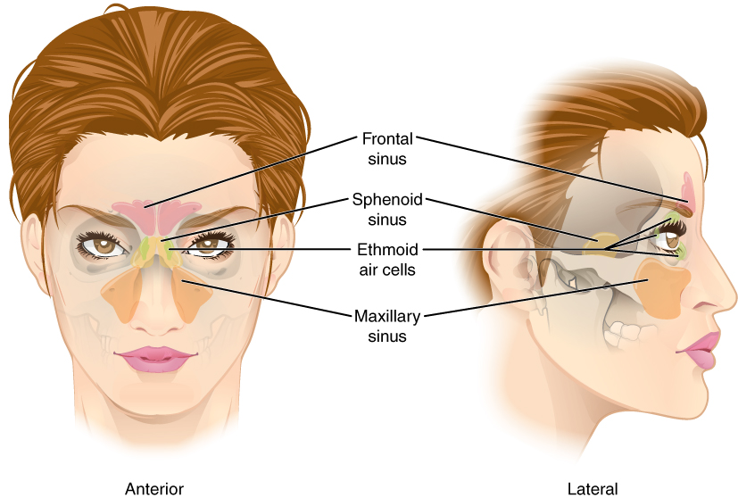
Paranasal sinuses

This figure shows a woman’s face and the location of the paranasal sinuses. The left panel shows the anterior view of the woman’s face with the sinuses labeled. The right panel shows the lateral view of the woman’s face with the same parts labeled.
The paranasal sinuses are hollow, air-filled spaces named for the skull bone that each occupies. The most anterior is the frontal sinus, located in the frontal bone above the eyebrows. The largest are the maxillary sinuses, located in the right and left maxillary bones below the orbits. The most posterior is the sphenoid sinus, located in the body of the sphenoid bone, under the sella turcica. The ethmoid air cells are multiple small spaces located in the right and left sides of the ethmoid bone, between the medial wall of the orbit and lateral wall of the upper nasal cavity.

Hyoid bone

The hyoid bone is an independent bone that does not contact any other bone and thus is not part of the skull ( [link] ). It is a small U-shaped bone located in the upper neck near the mandible, with the tips of the “U” pointing backwards. The hyoid serves as the base for the tongue above, and is attached to the larynx and the pharynx .
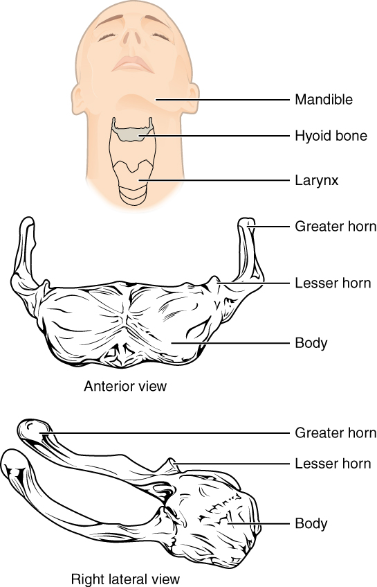
Hyoid bone

In this image, the location and structure of the hyoid bone are shown. The top panel shows a person’s face and neck, with the hyoid bone highlighted in grey. The middle panel shows the anterior view and the bottom panel shows the right anterior view.
The hyoid bone is located in the upper neck and does not join with any other bone. It provides attachments for muscles that act on the tongue, larynx, and pharynx.

References

Centers for Disease Control and Prevention (US). Injury prevention and control: traumatic brain injury [Internet]. Atlanta, GA; [cited 2013 Mar 18]. Available from: (External Link) .

Questions & Answers

A golfer on a fairway is 70 m away from the green, which sits below the level of the fairway by 20 m. If the golfer hits the ball at an angle of 40° with an initial speed of 20 m/s, how close to the green does she come?
Aislinn Reply
cm
tijani
what is titration
John Reply
what is physics
Siyaka Reply
A mouse of mass 200 g falls 100 m down a vertical mine shaft and lands at the bottom with a speed of 8.0 m/s. During its fall, how much work is done on the mouse by air resistance
Jude Reply
Can you compute that for me. Ty
Jude
what is the dimension formula of energy?
David Reply
what is viscosity?
David
what is inorganic
emma Reply
what is chemistry
Youesf Reply
what is inorganic
emma
Chemistry is a branch of science that deals with the study of matter,it composition,it structure and the changes it undergoes
Adjei
please, I'm a physics student and I need help in physics
Adjanou
chemistry could also be understood like the sexual attraction/repulsion of the male and female elements. the reaction varies depending on the energy differences of each given gender. + masculine -female.
Pedro
A ball is thrown straight up.it passes a 2.0m high window 7.50 m off the ground on it path up and takes 1.30 s to go past the window.what was the ball initial velocity
Krampah Reply
2. A sled plus passenger with total mass 50 kg is pulled 20 m across the snow (0.20) at constant velocity by a force directed 25° above the horizontal. Calculate (a) the work of the applied force, (b) the work of friction, and (c) the total work.
Sahid Reply
you have been hired as an espert witness in a court case involving an automobile accident. the accident involved car A of mass 1500kg which crashed into stationary car B of mass 1100kg. the driver of car A applied his brakes 15 m before he skidded and crashed into car B. after the collision, car A s
Samuel Reply
can someone explain to me, an ignorant high school student, why the trend of the graph doesn't follow the fact that the higher frequency a sound wave is, the more power it is, hence, making me think the phons output would follow this general trend?
Joseph Reply
Nevermind i just realied that the graph is the phons output for a person with normal hearing and not just the phons output of the sound waves power, I should read the entire thing next time
Joseph
Follow up question, does anyone know where I can find a graph that accuretly depicts the actual relative "power" output of sound over its frequency instead of just humans hearing
Joseph
"Generation of electrical energy from sound energy | IEEE Conference Publication | IEEE Xplore" ***ieeexplore.ieee.org/document/7150687?reload=true
Ryan
what's motion
Maurice Reply
what are the types of wave
Maurice
answer
Magreth
progressive wave
Magreth
hello friend how are you
Muhammad Reply
fine, how about you?
Mohammed
hi
Mujahid
A string is 3.00 m long with a mass of 5.00 g. The string is held taut with a tension of 500.00 N applied to the string. A pulse is sent down the string. How long does it take the pulse to travel the 3.00 m of the string?
yasuo Reply
Who can show me the full solution in this problem?
Reofrir Reply
Got questions? Join the online conversation and get instant answers!
Jobilize.com Reply

Get Jobilize Job Search Mobile App in your pocket Now!

Get it on Google Play Download on the App Store Now


Source:  OpenStax, Skeletal system. OpenStax CNX. Apr 17, 2015 Download for free at https://legacy.cnx.org/content/col11779/1.1
Google Play and the Google Play logo are trademarks of Google Inc.

Notification Switch

Would you like to follow the 'Skeletal system' conversation and receive update notifications?

Ask