<< Chapter < Page Chapter >> Page >

Learning objectives

  • Explain the role of microbial activity in diseases of the mouth and oral cavity
  • Compare the major characteristics of specific oral diseases and infections

Despite the presence of saliva and the mechanical forces of chewing and eating, some microbes thrive in the mouth. These microbes can cause damage to the teeth and can cause infections that have the potential to spread beyond the mouth and sometimes throughout the body.

Dental caries

Cavities of the teeth, known clinically as dental caries , are microbial lesions that cause damage to the teeth. Over time, the lesion can grow through the outer enamel layer to infect the underlying dentin or even the innermost pulp . If dental caries are not treated, the infection can become an abscess that spreads to the deeper tissues of the teeth , near the roots, or to the bloodstream.

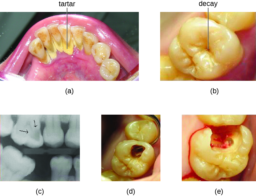
Tooth decay results from the metabolic activity of microbes that live on the teeth. A layer of proteins and carbohydrates forms when clean teeth come into contact with saliva. Microbes are attracted to this food source and form a biofilm called plaque . The most important cariogenic species in these biofilms is Streptococcus mutans . When sucrose , a disaccharide sugar from food, is broken down by bacteria in the mouth, glucose and fructose are produced. The glucose is used to make dextran , which is part of the extracellular matrix of the biofilm. Fructose is fermented, producing organic acids such as lactic acid . These acids dissolve the minerals of the tooth, including enamel , even though it is the hardest material in the body. The acids work even more quickly on exposed dentin ( [link] ). Over time, the plaque biofilm can become thick and eventually calcify. When a heavy plaque deposit becomes hardened in this way, it is called tartar or dental calculus ( [link] ). These substantial plaque biofilms can include a variety of bacterial species, including Streptococcus and Actinomyces species.
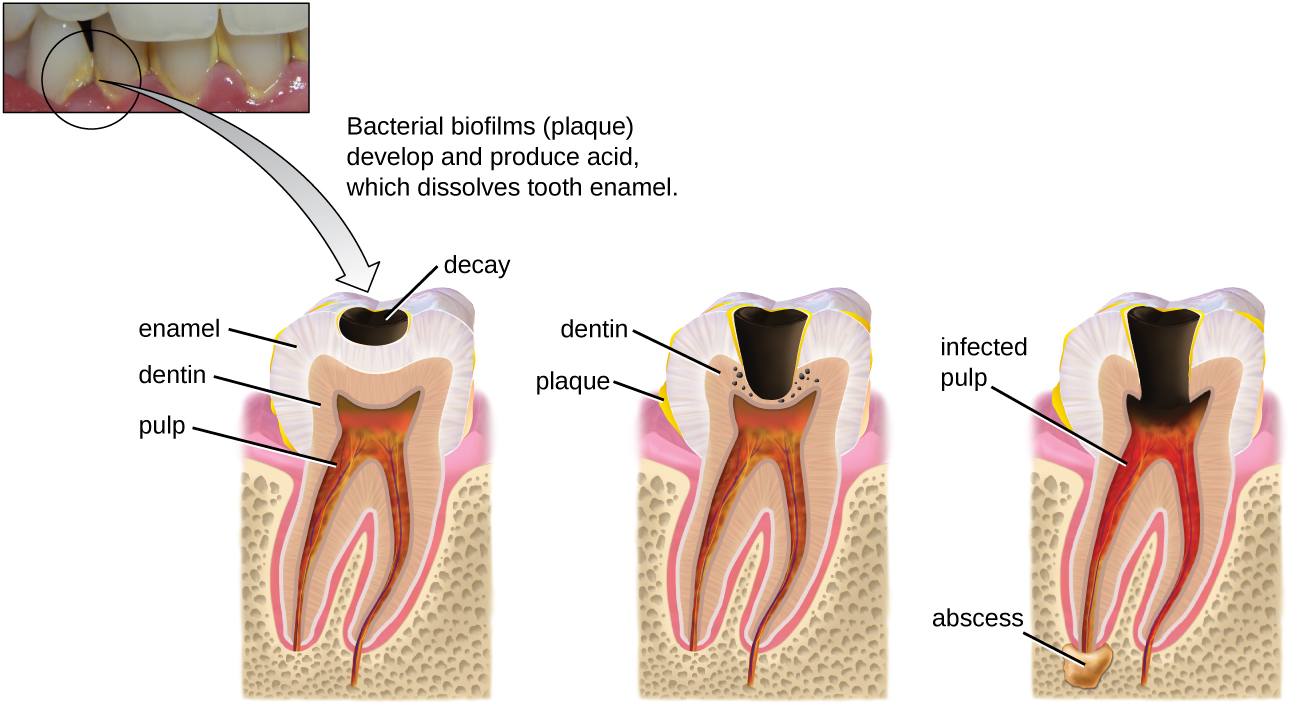
A photo of teeth with yellow plaque; label reads: bacterial biofilms (plaque) develop and produce acid which dissolves tooth enamel. This leads to a diagram showing the process. The first step show a black region labeled decay in the enamel; the dentin and pulp are not yet affected. Yellow material on the tooth and near the region of decay is labeled plaque. Next, the decay expands and is labeled abscess; this reaches the dentin layer. Finally, the abscess expands and causes an infected pulp.
Tooth decay occurs in stages. When bacterial biofilms (plaque) develop on teeth, the acids produced gradually dissolve the enamel, followed by the dentin. Eventually, if left untreated, the lesion may reach the pulp and cause an abscess. (credit: modification of work by “BruceBlaus”/Wikimedia Commons)
A) photo of a tooth with a dark spot labeled decay. B) micrograph of a tooth; dark regions have an arrow. C) photo of a tooth with a hole. D) photo of a tooth with a large, bleeding hole
(a) Tartar (dental calculus) is visible at the bases of these teeth. The darker deposits higher on the crowns are staining. (b) This tooth shows only a small amount of visible decay. (c) An X-ray of the same tooth shows that there is a dark area representing more decay inside the tooth. (d) Removal of a portion of the crown reveals the area of damage. (e) All of the cavity must be removed before filling. (credit: modification of work by “DRosenbach”/Wikimedia Commons)

Some tooth decay is visible from the outside, but it is not always possible to see all decay or the extent of the decay. X-ray imaging is used to produce radiographs that can be studied to look for deeper decay and damage to the root or bone ( [link] ). If not detected, the decay can reach the pulp or even spread to the bloodstream. Painful abscesses can develop.

Questions & Answers

describe the stage of eghopoisis
alupe Reply
what is a blood vessels
Sani Reply
what is plasma and is component
Fad Reply
what is the anterior
Tito Reply
Means front part of the body
Ibrahim
what is anatomy
Ruth Reply
describe the stage of ehopoisis
alupe
To better understand how the different part of the body works. To understand the physiology of the various structures in the body. To differentiate the systems of the human body .
Roseann Reply
what is hypogelersomia
aliyu Reply
what are the parts of the female reproductive system?
Orji Reply
what is anatomy
Divinefavour Reply
what are the six types of synovial joints and their ligaments
Darlington Reply
draw the six types of synovial joint and their ligaments
Darlington
System of human beings
Katumi Reply
System in humans body
Katumi
Diagram of animals and plants cell
Favour Reply
at what age does development of bone end
Alal Reply
how many bones are in the human upper layers
Daniel Reply
how many bones do we have
Nbeke
bones that form the wrist
Priscilla Reply
Got questions? Join the online conversation and get instant answers!
Jobilize.com Reply
Practice MCQ 4

Get Jobilize Job Search Mobile App in your pocket Now!

Get it on Google Play Download on the App Store Now


Source:  OpenStax, Microbiology. OpenStax CNX. Nov 01, 2016 Download for free at http://cnx.org/content/col12087/1.4
Google Play and the Google Play logo are trademarks of Google Inc.

Notification Switch

Would you like to follow the 'Microbiology' conversation and receive update notifications?

Ask