<< Chapter < Page Chapter >> Page >

Histology

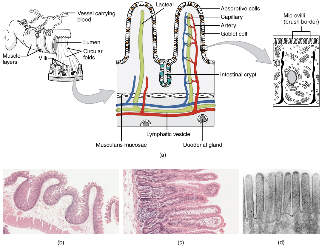
The wall of the small intestine is composed of the same four layers typically present in the alimentary system. However, three features of the mucosa and submucosa are unique. These features, which increase the absorptive surface area of the small intestine more than 600-fold, include circular folds, villi, and microvilli ( [link] ). These adaptations are most abundant in the first two-thirds of the small intestine, where the majority of absorption occurs.
Histology of the small intestine

Illustration (a) shows the histological cross-section of the small intestine. The left panel shows a small region of the small intestine, along with the blood vessels and the muscle layers. The middle panel shows a magnified view of a small region of the small intestine, highlighting the absorptive cells, the lacteal and the goblet cells. The right panel shows a further magnified view of the epithelial cells including the microvilli. Illustrations (b) shows a micrograph of the circular folds, and illustration (c) shows a micrograph of the villi. Illustration (d) shows an electron micrograph of the microvilli.
(a) The absorptive surface of the small intestine is vastly enlarged by the presence of circular folds, villi, and microvilli. (b) Micrograph of the circular folds. (c) Micrograph of the villi. (d) Electron micrograph of the microvilli. From left to right, LM x 56, LM x 508, EM x 196,000. (credit b-d: Micrograph provided by the Regents of University of Michigan Medical School © 2012)

Adaptations to increase surface area

There are three structural adaptations to the small intestine that increase the amount of area for food to be absorbed. A circular fold    is a deep ridge in the mucosa and submucosa. Beginning near the first part of the duodenum and ending near the middle of the ileum, these folds increase absorption. Their shape causes the chyme to spiral, rather than move in a straight line, through the small intestine. Spiraling slows the movement of chyme and provides the time needed for nutrients to be fully absorbed. Within the circular folds are small (0.5–1 mm long) hairlike projections called villi (singular = villus) that give the mucosa a furry texture. There are about 20 to 40 villi per square millimeter, increasing the surface area of the epithelium tremendously. Microvilli (singular = microvillus) are much smaller (1 µm) than villi. They are surface extensions of the plasma membrane of the mucosa’s epithelial cells. Although their small size makes it difficult to see each microvillus, their combined microscopic appearance suggests a mass of bristles, which is termed the brush border    . There are an estimated 200 million microvilli per square millimeter of small intestine, greatly expanding the surface area of the plasma membrane and thus greatly enhancing absorption.

Mechanical digestion in the small intestine

The movement of intestinal smooth muscles includes both segmentation and a form of peristalsis called migrating motility complexes. The kind of peristaltic mixing waves seen in the stomach are not observed here.

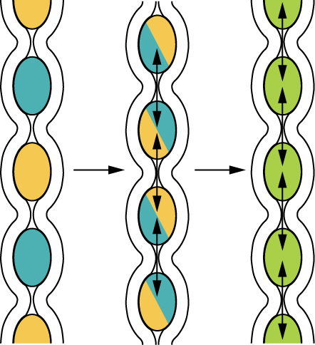
The smooth muscle layer of the small intestine is responsible for segmentation    .If you could see into the small intestine when it was going through segmentation, it would look as if the contents were being shoved incrementally back and forth. It combines the chyme with digestive juices and pushes food particles against the intestinal wall to be absorbed.
Segmentation

This diagram shows the process of segmentation in the intestines. The left panel shows the separation of chime, the middle panel shows the remixing of the chime by pushing it back together and the right panel indicates that the chime is being digested and absorbed.
Segmentation separates chyme and then pushes it back together, mixing it and providing time for digestion and absorption.

Chemical digestion in the small intestine

The digestion of proteins and carbohydrates, which partially occurs in the stomach, is completed in the small intestine with the aid of intestinal and pancreatic juices. Lipids arrive in the intestine largely undigested, so much of the focus here is on lipid digestion, which is facilitated by bile. Moreover, intestinal juice combines with pancreatic juice to provide a liquid medium that facilitates absorption. The intestine is also where most water is absorbed, via osmosis. The small intestine’s absorptive cells also produce digestive enzymes.

Questions & Answers

discuss how the following factors such as predation risk, competition and habitat structure influence animal's foraging behavior in essay form
Burnet Reply
location of cervical vertebra
KENNEDY Reply
What are acid
Sheriff Reply
define biology infour way
Happiness Reply
What are types of cell
Nansoh Reply
how can I get this book
Gatyin Reply
what is lump
Chineye Reply
what is cell
Maluak Reply
what is biology
Maluak
what's cornea?
Majak Reply
what are cell
Achol
Explain the following terms . (1) Abiotic factors in an ecosystem
Nomai Reply
Abiotic factors are non living components of ecosystem.These include physical and chemical elements like temperature,light,water,soil,air quality and oxygen etc
Qasim
Define the term Abiotic
Marial
what is biology
daniel Reply
what is diffusion
Emmanuel Reply
passive process of transport of low-molecular weight material according to its concentration gradient
AI-Robot
what is production?
Catherine
hello
Marial
Pathogens and diseases
how did the oxygen help a human being
Achol Reply
how did the nutrition help the plants
Achol Reply
Got questions? Join the online conversation and get instant answers!
Jobilize.com Reply

Get Jobilize Job Search Mobile App in your pocket Now!

Get it on Google Play Download on the App Store Now


Source:  OpenStax, Digestive system. OpenStax CNX. Feb 23, 2015 Download for free at http://legacy.cnx.org/content/col11761/1.1
Google Play and the Google Play logo are trademarks of Google Inc.

Notification Switch

Would you like to follow the 'Digestive system' conversation and receive update notifications?

Ask