| << Chapter < Page | Chapter >> Page > |

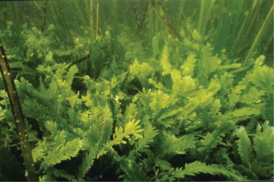
True multicellular organisms, such as the sea lettuce, Ulva , are represented among the chlorophytes. In addition, some chlorophytes exist as large, multinucleate, single cells. Species in the genus Caulerpa exhibit flattened fern-like foliage and can reach lengths of 3 meters ( [link] ). Caulerpa species undergo nuclear division, but their cells do not complete cytokinesis, remaining instead as massive and elaborate single cells.

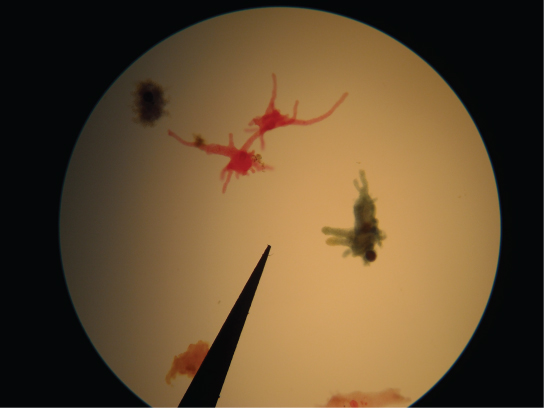
The amoebozoans characteristically exhibit pseudopodia that extend like tubes or flat lobes, rather than the hair-like pseudopodia of rhizarian amoeba ( [link] ). The Amoebozoa include several groups of unicellular amoeba-like organisms that are free-living or parasites.

A subset of the amoebozoans, the slime molds, has several morphological similarities to fungi that are thought to be the result of convergent evolution. For instance, during times of stress, some slime molds develop into spore-generating fruiting bodies, much like fungi.
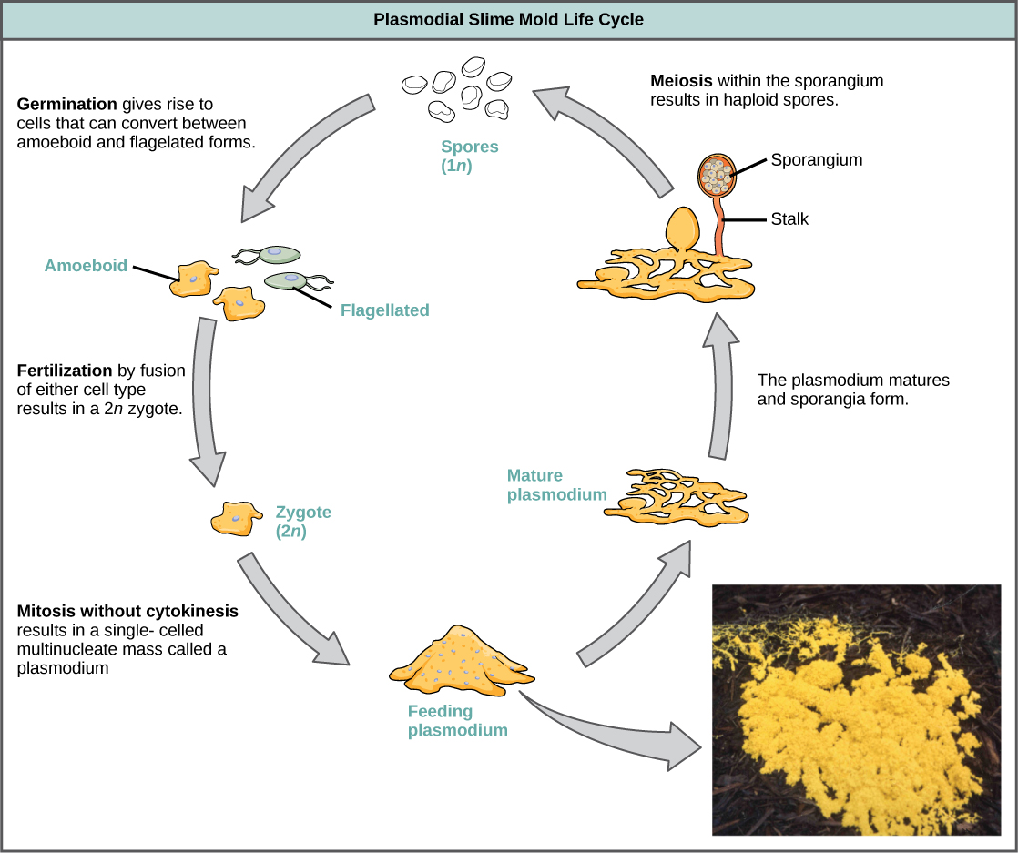
The slime molds are categorized on the basis of their life cycles into plasmodial or cellular types. Plasmodial slime molds are composed of large, multinucleate cells and move along surfaces like an amorphous blob of slime during their feeding stage ( [link] ). Food particles are lifted and engulfed into the slime mold as it glides along. Upon maturation, the plasmodium takes on a net-like appearance with the ability to form fruiting bodies, or sporangia, during times of stress. Haploid spores are produced by meiosis within the sporangia, and spores can be disseminated through the air or water to potentially land in more favorable environments. If this occurs, the spores germinate to form ameboid or flagellate haploid cells that can combine with each other and produce a diploid zygotic slime mold to complete the life cycle.

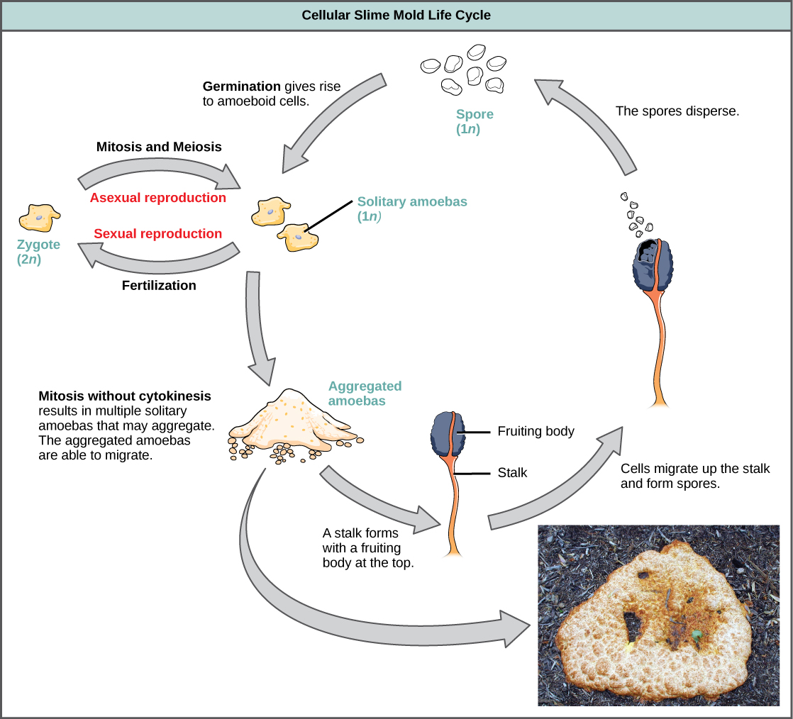
The cellular slime molds function as independent amoeboid cells when nutrients are abundant ( [link] ). When food is depleted, cellular slime molds pile onto each other into a mass of cells that behaves as a single unit, called a slug. Some cells in the slug contribute to a 2–3-millimeter stalk, drying up and dying in the process. Cells atop the stalk form an asexual fruiting body that contains haploid spores. As with plasmodial slime molds, the spores are disseminated and can germinate if they land in a moist environment. One representative genus of the cellular slime molds is Dictyostelium , which commonly exists in the damp soil of forests.

View this site to see the formation of a fruiting body by a cellular slime mold.
The opisthokonts include the animal-like choanoflagellates, which are believed to resemble the common ancestor of sponges and, in fact, all animals. Choanoflagellates include unicellular and colonial forms, and number about 244 described species. These organisms exhibit a single, apical flagellum that is surrounded by a contractile collar composed of microvilli. The collar uses a similar mechanism to sponges to filter out bacteria for ingestion by the protist. The morphology of choanoflagellates was recognized early on as resembling the collar cells of sponges, and suggesting a possible relationship to animals.
The Mesomycetozoa form a small group of parasites, primarily of fish, and at least one form that can parasitize humans. Their life cycles are poorly understood. These organisms are of special interest, because they appear to be so closely related to animals. In the past, they were grouped with fungi and other protists based on their morphology.
The process of classifying protists into meaningful groups is ongoing, but genetic data in the past 20 years have clarified many relationships that were previously unclear or mistaken. The majority view at present is to order all eukaryotes into six supergroups: Excavata, Chromalveolata, Rhizaria, Archaeplastida, Amoebozoa, and Opisthokonta. The goal of this classification scheme is to create clusters of species that all are derived from a common ancestor. At present, the monophyly of some of the supergroups are better supported by genetic data than others. Although tremendous variation exists within the supergroups, commonalities at the morphological, physiological, and ecological levels can be identified.
[link] Which of the following statements about Paramecium sexual reproduction is false?
[link] C
[link] Which of the following statements about the Laminaria life cycle is false?
[link] C

Notification Switch
Would you like to follow the 'Biology' conversation and receive update notifications?