<< Chapter < Page Chapter >> Page >

Capillary network within the nephron

The capillary network that originates from the renal arteries supplies the nephron with blood that needs to be filtered. The branch that enters the glomerulus is called the afferent arteriole    . The branch that exits the glomerulus is called the efferent arteriole    . Within the glomerulus, the network of capillaries is called the glomerular capillary bed. Once the efferent arteriole exits the glomerulus, it forms the peritubular capillary network    , which surrounds and interacts with parts of the renal tubule. In cortical nephrons, the peritubular capillary network surrounds the PCT and DCT. In juxtamedullary nephrons, the peritubular capillary network forms a network around the loop of Henle and is called the vasa recta    .

Go to this website to see another coronal section of the kidney and to explore an animation of the workings of nephrons.

Kidney function and physiology

Kidneys filter blood in a three-step process. First, the nephrons filter blood that runs through the capillary network in the glomerulus. Almost all solutes, except for proteins, are filtered out into the glomerulus by a process called glomerular filtration    . Second, the filtrate is collected in the renal tubules. Most of the solutes get reabsorbed in the PCT by a process called tubular reabsorption    . In the loop of Henle, the filtrate continues to exchange solutes and water with the renal medulla and the peritubular capillary network. Water is also reabsorbed during this step. Then, additional solutes and wastes are secreted into the kidney tubules during tubular secretion    , which is, in essence, the opposite process to tubular reabsorption. The collecting ducts collect filtrate coming from the nephrons and fuse in the medullary papillae. From here, the papillae deliver the filtrate, now called urine, into the minor calyces that eventually connect to the ureters through the renal pelvis. This entire process is illustrated in [link] .
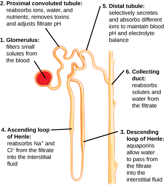
Illustration labels parts of a nephron and their function. The nephron begins at the glomerulus, a spherical structure that filters small solutes from the blood. The filtrate then enters a winding proximal convoluted tubule, which reabsorbs ions, water, and nutrients, and removes toxins and adjusts the filtrate pH. The proximal convoluted tubule empties into the descending loop of Henle. Aquaporins in the descending loop allow water to pass from the filtrate to the interstitial fluid. The descending loop of Henle turns into the ascending loop of Henle. Both the descending loop and ascending loop are thin at the bottom, and turn thick about a third of the way up. In the ascending loop of Henle, sodium and chlorine ions are reabsorbed from the filtrate into the interstitial fluid. The ascending loop of Henle empties into the distal convoluted tubule, which selectively secretes and absorbs ions to maintain blood pH and electrolyte balance. The distal convoluted tubule empties into a collecting duct, which reabsorbs water and solutes from the filtrate. The collecting duct travels down, toward the middle of the kidney.
Each part of the nephron performs a different function in filtering waste and maintaining homeostatic balance. (1) The glomerulus forces small solutes out of the blood by pressure. (2) The proximal convoluted tubule reabsorbs ions, water, and nutrients from the filtrate into the interstitial fluid, and actively transports toxins and drugs from the interstitial fluid into the filtrate. The proximal convoluted tubule also adjusts blood pH by selectively secreting ammonia (NH 3 ) into the filtrate, where it reacts with H + to form NH 4 + . The more acidic the filtrate, the more ammonia is secreted. (3) The descending loop of Henle is lined with cells containing aquaporins that allow water to pass from the filtrate into the interstitial fluid. (4) In the thin part of the ascending loop of Henle, Na + and Cl - ions diffuse into the interstitial fluid. In the thick part, these same ions are actively transported into the interstitial fluid. Because salt but not water is lost, the filtrate becomes more dilute as it travels up the limb. (5) In the distal convoluted tubule, K + and H + ions are selectively secreted into the filtrate, while Na + , Cl - , and HCO 3 - ions are reabsorbed to maintain pH and electrolyte balance in the blood. (6) The collecting duct reabsorbs solutes and water from the filtrate, forming dilute urine. (credit: modification of work by NIDDK)

Questions & Answers

1. Discuss the processes involved during exchange of fluids between intra and extracellular space.
Mustapha Reply
what are components of cells
ofosola Reply
twugzfisfjxxkvdsifgfuy7 it
Sami
58214993
Sami
what is a salt
John
the difference between male and female reproduction
John
what is computed
IBRAHIM Reply
what is biology
IBRAHIM
what is the full meaning of biology
IBRAHIM
what is biology
Jeneba
what is cell
Kuot
425844168
Sami
what is biology
Inenevwo
what is cytoplasm
Emmanuel Reply
structure of an animal cell
Arrey Reply
what happens when the eustachian tube is blocked
Puseletso Reply
what's atoms
Achol Reply
discuss how the following factors such as predation risk, competition and habitat structure influence animal's foraging behavior in essay form
Burnet Reply
cell?
Kuot
location of cervical vertebra
KENNEDY Reply
What are acid
Sheriff Reply
define biology infour way
Happiness Reply
What are types of cell
Nansoh Reply
how can I get this book
Gatyin Reply
what is lump
Chineye Reply
what is cell
Maluak Reply
what is biology
Maluak
what is vertibrate
Jeneba

Get Jobilize Job Search Mobile App in your pocket Now!

Get it on Google Play Download on the App Store Now


Source:  OpenStax, Biology. OpenStax CNX. Feb 29, 2016 Download for free at http://cnx.org/content/col11448/1.10
Google Play and the Google Play logo are trademarks of Google Inc.

Notification Switch

Would you like to follow the 'Biology' conversation and receive update notifications?

Ask