<< Chapter < Page Chapter >> Page >
Figure A is the standing-wave pattern for a string clamped in the wall. The distance between each node corresponds to the half gamma. Figure B is the standing-wave pattern for an electron wave trapped in the third Bohr orbit in the hydrogen atom. The wave has a circular shape with six nodes. The distance between each two node corresponds to the gamma.
Standing-wave pattern: (a) a stretched string clamped at the walls; (b) an electron wave trapped in the third Bohr orbit in the hydrogen atom.

The electron wave in the ground state of hydrogen

Find the de Broglie wavelength of an electron in the ground state of hydrogen.

Strategy

We combine the first quantization condition in [link] with [link] and use [link] for the first Bohr radius with n = 1 .

Solution

When n = 1 and r n = a 0 = 0.529 Å, the Bohr quantization condition gives a 0 p = 1 · p = / a 0 . The electron wavelength is:

λ = h / p = h / / a 0 = 2 π a 0 = 2 π ( 0.529 Å ) = 3.324 Å .

Significance

We obtain the same result when we use [link] directly.

Got questions? Get instant answers now!

Check Your Understanding Find the de Broglie wavelength of an electron in the third excited state of hydrogen.

λ = 2 π n a 0 = 2 ( 3.324 Å ) = 6.648 Å

Got questions? Get instant answers now!

Experimental confirmation of matter waves came in 1927 when C. Davisson and L. Germer performed a series of electron-scattering experiments that clearly showed that electrons do behave like waves. Davisson and Germer did not set up their experiment to confirm de Broglie’s hypothesis: The confirmation came as a byproduct of their routine experimental studies of metal surfaces under electron bombardment.

In the particular experiment that provided the very first evidence of electron waves (known today as the Davisson–Germer experiment    ), they studied a surface of nickel. Their nickel sample was specially prepared in a high-temperature oven to change its usual polycrystalline structure to a form in which large single-crystal domains occupy the volume. [link] shows the experimental setup. Thermal electrons are released from a heated element (usually made of tungsten) in the electron gun and accelerated through a potential difference Δ V , becoming a well-collimated beam of electrons produced by an electron gun. The kinetic energy K of the electrons is adjusted by selecting a value of the potential difference in the electron gun. This produces a beam of electrons with a set value of linear momentum, in accordance with the conservation of energy:

e Δ V = K = p 2 2 m p = 2 m e Δ V .

The electron beam is incident on the nickel sample in the direction normal to its surface. At the surface, it scatters in various directions. The intensity of the beam scattered in a selected direction φ is measured by a highly sensitive detector. The detector’s angular position with respect to the direction of the incident beam can be varied from φ = 0 ° to φ = 90 ° . The entire setup is enclosed in a vacuum chamber to prevent electron collisions with air molecules, as such thermal collisions would change the electrons’ kinetic energy and are not desirable.
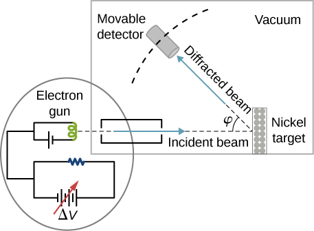
Figure shows the schematics of the experimental setup of the Davisson–Germer diffraction experiment. A beam of electrons is emitted by the electron gun, passes through the collimator, and hits Nickel target. Diffracted beam forms an angle phi with the incident beam and is detected by a moving detector. All of this is shown happening in a vacuum
Schematics of the experimental setup of the Davisson–Germer diffraction experiment. A well-collimated beam of electrons is scattered off the nickel target. The kinetic energy of electrons in the incident beam is selected by adjusting a variable potential, Δ V , in the electron gun. Intensity of the scattered electron beam is measured for a range of scattering angles φ , whereas the distance between the detector and the target does not change.

When the nickel target has a polycrystalline form with many randomly oriented microscopic crystals, the incident electrons scatter off its surface in various random directions. As a result, the intensity of the scattered electron beam is much the same in any direction, resembling a diffuse reflection of light from a porous surface. However, when the nickel target has a regular crystalline structure, the intensity of the scattered electron beam shows a clear maximum at a specific angle and the results show a clear diffraction pattern (see [link] ). Similar diffraction patterns formed by X-rays scattered by various crystalline solids were studied in 1912 by father-and-son physicists William H. Bragg and William L. Bragg . The Bragg law in X-ray crystallography provides a connection between the wavelength λ of the radiation incident on a crystalline lattice, the lattice spacing, and the position of the interference maximum in the diffracted radiation (see Diffraction ).

Questions & Answers

what are components of cells
ofosola Reply
twugzfisfjxxkvdsifgfuy7 it
Sami
58214993
Sami
what is a salt
John
the difference between male and female reproduction
John
what is computed
IBRAHIM Reply
what is biology
IBRAHIM
what is the full meaning of biology
IBRAHIM
what is biology
Jeneba
what is cell
Kuot
425844168
Sami
what is cytoplasm
Emmanuel Reply
structure of an animal cell
Arrey Reply
what happens when the eustachian tube is blocked
Puseletso Reply
what's atoms
Achol Reply
discuss how the following factors such as predation risk, competition and habitat structure influence animal's foraging behavior in essay form
Burnet Reply
cell?
Kuot
location of cervical vertebra
KENNEDY Reply
What are acid
Sheriff Reply
define biology infour way
Happiness Reply
What are types of cell
Nansoh Reply
how can I get this book
Gatyin Reply
what is lump
Chineye Reply
what is cell
Maluak Reply
what is biology
Maluak
what is vertibrate
Jeneba
what's cornea?
Majak Reply
what are cell
Achol
Got questions? Join the online conversation and get instant answers!
Jobilize.com Reply
Practice Key Terms 5

Get Jobilize Job Search Mobile App in your pocket Now!

Get it on Google Play Download on the App Store Now


Source:  OpenStax, University physics volume 3. OpenStax CNX. Nov 04, 2016 Download for free at http://cnx.org/content/col12067/1.4
Google Play and the Google Play logo are trademarks of Google Inc.

Notification Switch

Would you like to follow the 'University physics volume 3' conversation and receive update notifications?

Ask