<< Chapter < Page Chapter >> Page >
By the end of this section, you will be able to:
  • Explain the physics behind the operation of microscopes and telescopes
  • Describe the image created by these instruments and calculate their magnifications

Microscopes and telescopes are major instruments that have contributed hugely to our current understanding of the micro- and macroscopic worlds. The invention of these devices led to numerous discoveries in disciplines such as physics, astronomy, and biology, to name a few. In this section, we explain the basic physics that make these instruments work.

Microscopes

Although the eye is marvelous in its ability to see objects large and small, it obviously is limited in the smallest details it can detect. The desire to see beyond what is possible with the naked eye led to the use of optical instruments. We have seen that a simple convex lens can create a magnified image, but it is hard to get large magnification with such a lens. A magnification greater than 5 × is difficult without distorting the image. To get higher magnification, we can combine the simple magnifying glass with one or more additional lenses. In this section, we examine microscopes that enlarge the details that we cannot see with the naked eye.

Microscopes were first developed in the early 1600s by eyeglass makers in The Netherlands and Denmark. The simplest compound microscope    is constructed from two convex lenses ( [link] ). The objective    lens is a convex lens of short focal length (i.e., high power) with typical magnification from 5 × to 100 × . The eyepiece    , also referred to as the ocular, is a convex lens of longer focal length.

The purpose of a microscope is to create magnified images of small objects, and both lenses contribute to the final magnification. Also, the final enlarged image is produced sufficiently far from the observer to be easily viewed, since the eye cannot focus on objects or images that are too close (i.e., closer than the near point of the eye).
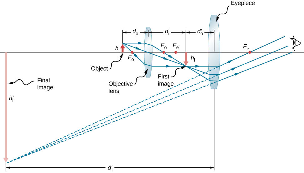
Figure shows from left to right: an object with height h, a bi-convex lens labeled objective lens at a distance d subscript o from the object, an inverted image with height h subscript i labeled first image at a distance d subscript i from the objective lens, a bi-convex lens labeled eyepiece at a distance d subscript o prime from the first image and finally the eye of the observer. Rays originate from the top of the object and pass through the objective lens to converge at the top of the inverted image. They travel further and enter the eyepiece, from where they deviate to reach the eye. The back extensions of the deviated rays converge at the tip of a much larger inverted image to the far left of the figure. The height of this image is h subscript i prime and its distance from the eyepiece is d subscript i prime.
A compound microscope is composed of two lenses: an objective and an eyepiece. The objective forms the first image, which is larger than the object. This first image is inside the focal length of the eyepiece and serves as the object for the eyepiece. The eyepiece forms final image that is further magnified.

To see how the microscope in [link] forms an image, consider its two lenses in succession. The object is just beyond the focal length f obj of the objective lens, producing a real, inverted image that is larger than the object. This first image serves as the object for the second lens, or eyepiece. The eyepiece is positioned so that the first image is within its focal length f eye , so that it can further magnify the image. In a sense, it acts as a magnifying glass that magnifies the intermediate image produced by the objective. The image produced by the eyepiece is a magnified virtual image. The final image remains inverted but is farther from the observer than the object, making it easy to view.

The eye views the virtual image created by the eyepiece, which serves as the object for the lens in the eye. The virtual image formed by the eyepiece is well outside the focal length of the eye, so the eye forms a real image on the retina.

Questions & Answers

what's atoms
Achol Reply
discuss how the following factors such as predation risk, competition and habitat structure influence animal's foraging behavior in essay form
Burnet Reply
location of cervical vertebra
KENNEDY Reply
What are acid
Sheriff Reply
define biology infour way
Happiness Reply
What are types of cell
Nansoh Reply
how can I get this book
Gatyin Reply
what is lump
Chineye Reply
what is cell
Maluak Reply
what is biology
Maluak
what's cornea?
Majak Reply
what are cell
Achol
Explain the following terms . (1) Abiotic factors in an ecosystem
Nomai Reply
Abiotic factors are non living components of ecosystem.These include physical and chemical elements like temperature,light,water,soil,air quality and oxygen etc
Qasim
Define the term Abiotic
Marial
what is biology
daniel Reply
what is diffusion
Emmanuel Reply
passive process of transport of low-molecular weight material according to its concentration gradient
AI-Robot
what is production?
Catherine
hello
Marial
Pathogens and diseases
how did the oxygen help a human being
Achol Reply
Got questions? Join the online conversation and get instant answers!
Jobilize.com Reply
Practice Key Terms 6

Get Jobilize Job Search Mobile App in your pocket Now!

Get it on Google Play Download on the App Store Now


Source:  OpenStax, University physics volume 3. OpenStax CNX. Nov 04, 2016 Download for free at http://cnx.org/content/col12067/1.4
Google Play and the Google Play logo are trademarks of Google Inc.

Notification Switch

Would you like to follow the 'University physics volume 3' conversation and receive update notifications?

Ask