| << Chapter < Page | Chapter >> Page > |

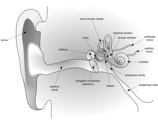
The human ear is divided into three main sections: the outer, middle, and inner ear. Let's follow the journey of a sound wave from the pinna (outermost part) to the auditory nerve (innermost part) which transmits a signal to the brain. The pinna is the part of the ear we typically think of when we refer to the ear. Its mainfunction is to collect and focus an incident sound wave. The wave then travels through the ear canal until it meets the eardrum. Thepressure fluctuations of the sound wave make the eardrum vibrate. The three very small bones of the middle ear, the malleus (hammer),the incus (anvil), and the stapes (stirrup), transmit the signal through to the elliptical window. The elliptical window is the beginning of theinner ear. From the elliptical window the sound waves are transmitted through the liquid in the inner ear and interpreted as sounds by the brain.The inner ear, made of the semicircular canals, the cochlea, and the auditory nerve, is filled with fluid. The fluid allows the body todetect quick movements and maintain balance. The snail-shaped cochlea is covered in nerve cells. There are more than 25 000 hairlikenerve cells. Different nerve cells vibrate with different frequencies. When a nerve cell vibrates, it releases electrical impulsesto the auditory nerve. The impulses are sent to the brain through the auditory nerve and understood as sound.
Ultrasound is sound with a frequency that is higher than 20 kHz. Some animals, such as dogs, dolphins, and bats, have an upper limit that is greater than that of the human ear and can hear ultrasound.
| Application | Lowest Frequency (kHz) | Highest Frequency (kHz) |
| Cleaning (e.g. Jewelery) | 20 | 40 |
| Material testing for flaws | 50 | 500 |
| Welding of plastics | 15 | 40 |
| Tumour ablation | 250 | 2000 |
The most common use of ultrasound is to create images, and has industrial and medical applications. The use of ultrasound to create images is based on the reflection and transmission of a wave at a boundary. When an ultrasound wave travels inside an object that is made up of different materials such as the human body, each time it encounters a boundary, e.g. between bone and muscle, or muscle and fat, part of the wave is reflected and part of it is transmitted. The reflected rays are detected and used to construct an image of the object.
Ultrasound in medicine can visualise muscle and soft tissue, making them useful for scanning the organs, and is commonly used during pregnancy. Ultrasound is a safe, non-invasive method of looking inside the human body.
Ultrasound sources may be used to generate local heating in biological tissue, with applications in physical therapy and cancer treatment. Focussed ultrasound sources may be used to break up kidney stones.
Ultrasonic cleaners, sometimes called supersonic cleaners, are used at frequencies from 20-40 kHz for jewellery, lenses and other optical parts, watches, dental instruments, surgical instruments and industrial parts. These cleaners consist of containers with a fluid in which the object to be cleaned is placed. Ultrasonic waves are then sent into the fluid. The main mechanism for cleaning action in an ultrasonic cleaner is actually the energy released from the collapse of millions of microscopic bubbles occurring in the liquid of the cleaner.

Notification Switch
Would you like to follow the 'Siyavula textbooks: grade 10 physical science [caps]' conversation and receive update notifications?