| << Chapter < Page | Chapter >> Page > |
The skeletal muscles are divided into axial (muscles of the trunk and head) and appendicular (muscles of the arms and legs) categories. This system reflects the bones of the skeleton system, which are also arranged in this manner. The axial muscles are grouped based on location, function, or both. Some of the axial muscles may seem to blur the boundaries because they cross over to the appendicular skeleton. The first grouping of the axial muscles you will review includes the muscles of the head and neck, then you will review the muscles of the vertebral column, and finally you will review the oblique and rectus muscles.
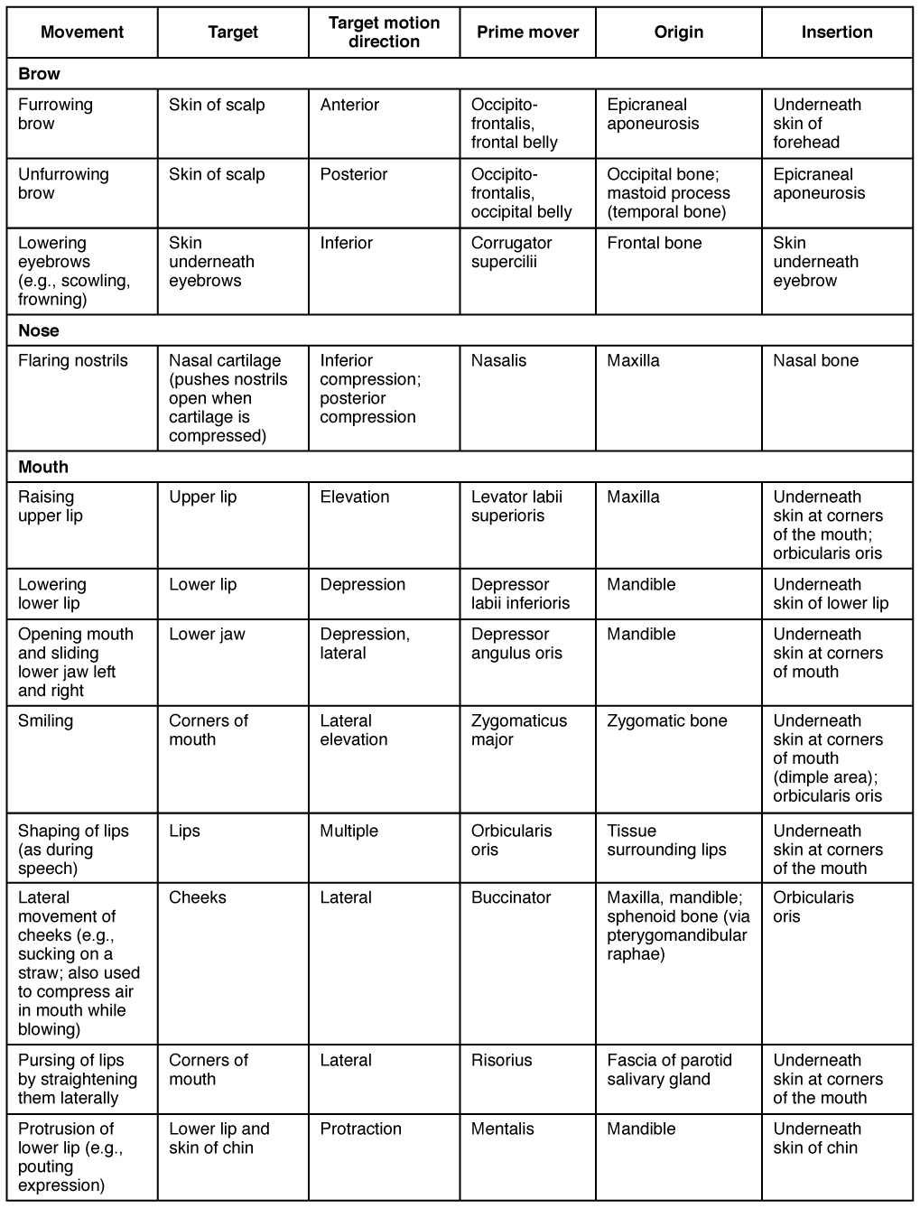
The origins of the muscles of facial expression are on the surface of the skull (remember, the origin of a muscle does not move). The insertions of these muscles have fibers intertwined with connective tissue and the dermis of the skin. Because the muscles insert in the skin rather than on bone, when they contract, the skin moves to create facial expression ( [link] ).

The orbicularis oris is a circular muscle that moves the lips, and the orbicularis oculi is a circular muscle that closes the eye. The occipitofrontalis muscle moves up the scalp and eyebrows. The muscle has a frontal belly and an occipital (near the occipital bone on the posterior part of the skull) belly. In other words, there is a muscle on the forehead ( frontalis ) and one on the back of the head ( occipitalis ), but there is no muscle across the top of the head. Instead, the two bellies are connected by a broad tendon called the epicranial aponeurosis , or galea aponeurosis (galea = “apple”). The physicians originally studying human anatomy thought the skull looked like an apple.
The majority of the face is composed of the buccinator muscle, which compresses the cheek. This muscle allows you to whistle, blow, and suck; and it contributes to the action of chewing. There are several small facial muscles, one of which is the corrugator supercilii , which is the prime mover of the eyebrows. Place your finger on your eyebrows at the point of the bridge of the nose. Raise your eyebrows as if you were surprised and lower your eyebrows as if you were frowning. With these movements, you can feel the action of the corrugator supercilli. Additional muscles of facial expression are presented in [link] .

The movement of the eyeball is under the control of the extrinsic eye muscles , which originate outside the eye and insert onto the outer surface of the white of the eye. These muscles are located inside the eye socket and cannot be seen on any part of the visible eyeball ( [link] and [link] ). If you have ever been to a doctor who held up a finger and asked you to follow it up, down, and to both sides, he or she is checking to make sure your eye muscles are acting in a coordinated pattern.

Notification Switch
Would you like to follow the 'Anatomy & Physiology: support and movement' conversation and receive update notifications?