<< Chapter < Page Chapter >> Page >
By the end of this section, you will be able to:
  • Describe the basic structure of a typical prokaryote
  • Describe important differences in structure between Archaea and Bacteria

There are many differences between prokaryotic and eukaryotic cells. However, all cells have four common structures: the plasma membrane, which functions as a barrier for the cell and separates the cell from its environment; the cytoplasm, a jelly-like substance inside the cell; nucleic acids, the genetic material of the cell; and ribosomes, where protein synthesis takes place. Prokaryotes come in various shapes, but many fall into three categories: cocci (spherical), bacilli (rod-shaped), and spirilli (spiral-shaped) ( [link] ).
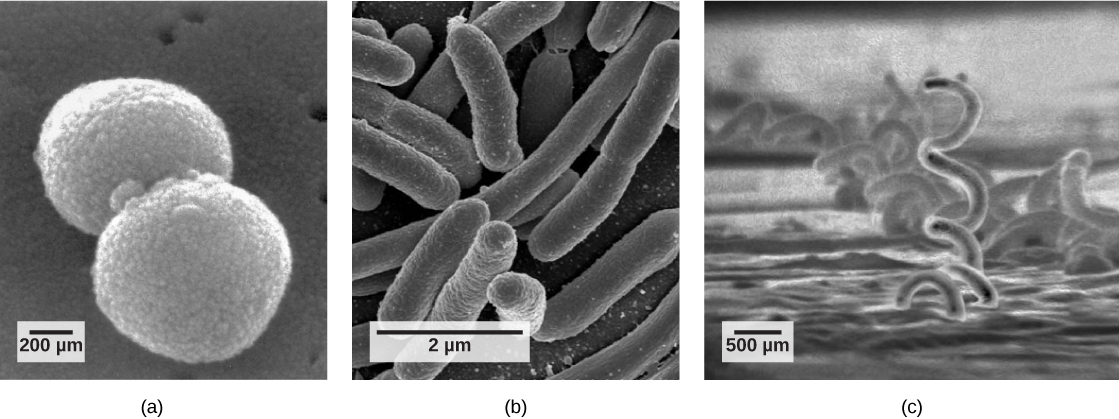
Part a: The micrograph shows ball-shaped cocci about 0.9 microns long. Part b: The micrograph shows hotdog-shaped bacilli about 2 microns long. Part c: The micrograph shows corkscrew-shaped spirilli that are quite long and 2 microns in diameter.
Prokaryotes fall into three basic categories based on their shape, visualized here using scanning electron microscopy: (a) cocci, or spherical (a pair is shown); (b) bacilli, or rod-shaped; and (c) spirilli, or spiral-shaped. (credit a: modification of work by Janice Haney Carr, Dr. Richard Facklam, CDC; credit c: modification of work by Dr. David Cox; scale-bar data from Matt Russell)

The prokaryotic cell

Recall that prokaryotes ( [link] ) are unicellular organisms that lack organelles or other internal membrane-bound structures. Therefore, they do not have a nucleus but instead generally have a single chromosome—a piece of circular, double-stranded DNA located in an area of the cell called the nucleoid. Most prokaryotes have a cell wall outside the plasma membrane.
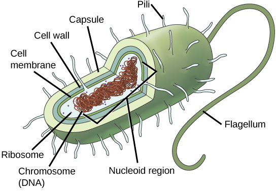
In this illustration, the prokaryotic cell is rod shaped. The circular chromosome is concentrated in a region called the nucleoid. The fluid inside the cell is called the cytoplasm. Ribosomes, depicted as small circles, float in the cytoplasm. The cytoplasm is encased by a plasma membrane, which in turn is encased by a cell wall. A capsule surrounds the cell wall. The bacterium depicted has a flagellum protruding from one narrow end. Pili are small protrusions that project from the capsule all over the bacterium, like hair.
The features of a typical prokaryotic cell are shown.

Recall that prokaryotes are divided into two different domains, Bacteria and Archaea, which together with Eukarya, comprise the three domains of life ( [link] ).
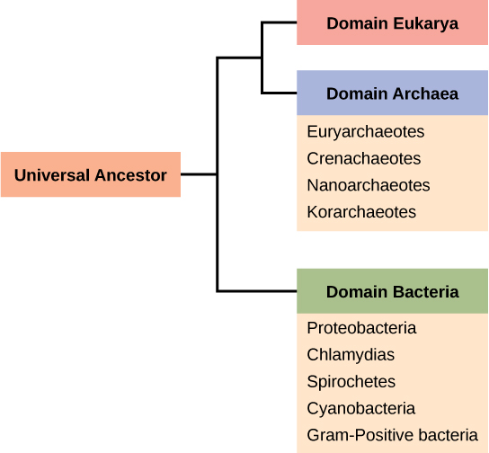
 The trunk of the phylogenetic tree is a universal ancestor. The tree forms two branches. One branch leads to the domain bacteria, which includes the phyla proteobacteria, chlamydias, spirochetes, cyanobacteria, and Gram-positive bacteria. The other branch branches again, into the eukarya and archaea domains. Domain archaea includes the phyla euryarchaeotes, crenarchaeotes, nanoarchaeotes, and korarchaeotea.
Bacteria and Archaea are both prokaryotes but differ enough to be placed in separate domains. An ancestor of modern Archaea is believed to have given rise to Eukarya, the third domain of life. Archaeal and bacterial phyla are shown; the evolutionary relationship between these phyla is still open to debate.

The composition of the cell wall differs significantly between the domains Bacteria and Archaea. The composition of their cell walls also differs from the eukaryotic cell walls found in plants (cellulose) or fungi and insects (chitin). The cell wall functions as a protective layer, and it is responsible for the organism’s shape. Some bacteria have an outer capsule    outside the cell wall. Other structures are present in some prokaryotic species, but not in others ( [link] ). For example, the capsule found in some species enables the organism to attach to surfaces, protects it from dehydration and attack by phagocytic cells, and makes pathogens more resistant to our immune responses. Some species also have flagella (singular, flagellum) used for locomotion, and pili (singular, pilus) used for attachment to surfaces. Plasmids, which consist of extra-chromosomal DNA, are also present in many species of bacteria and archaea.

Characteristics of phyla of Bacteria are described in [link] and [link] ; Archaea are described in [link] .
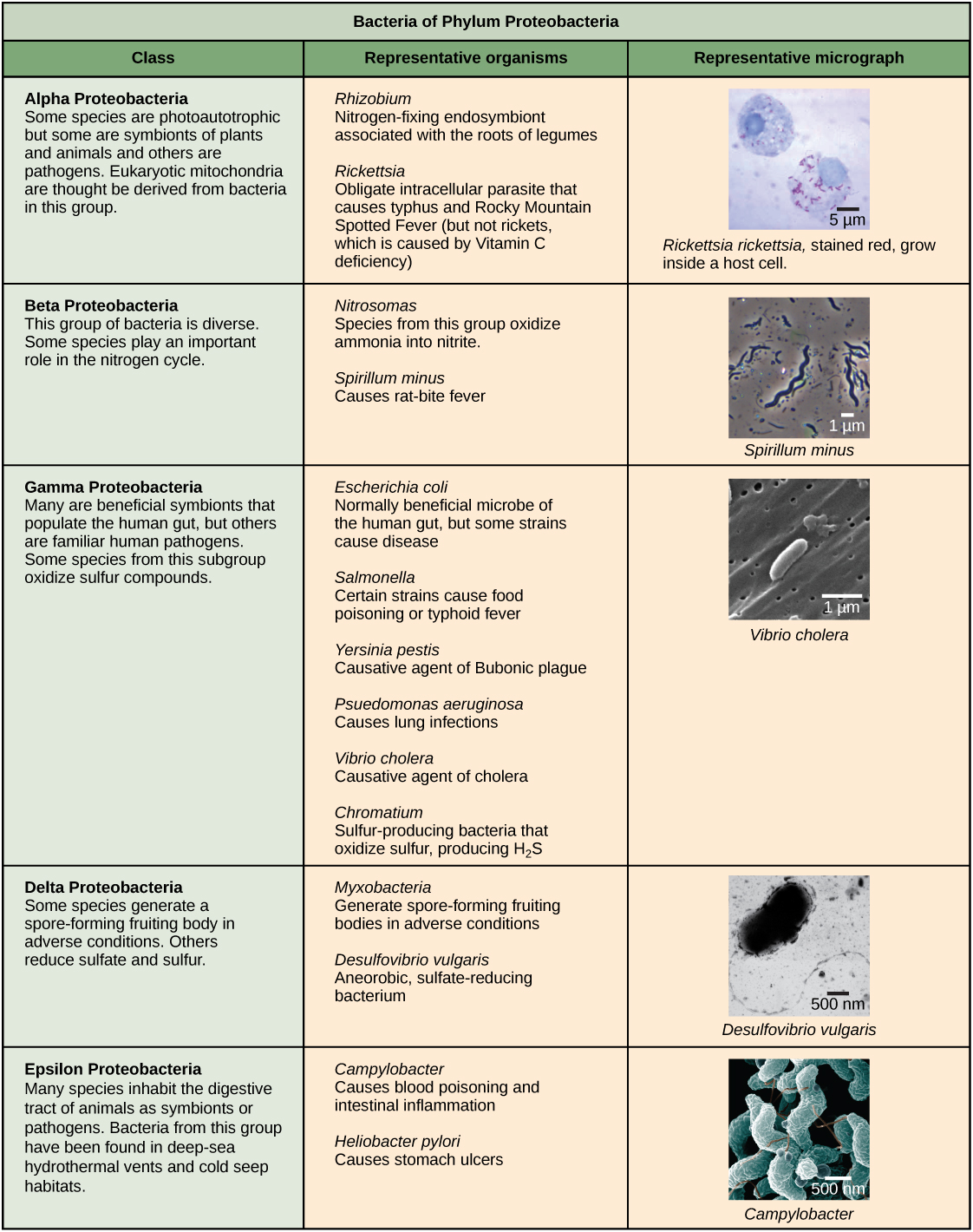
Characteristics of the five phyla of bacteria are described. The first phylum described is proteobacteria, which includes five classes, alpha, beta, gamma, delta and epsilon. Most species of Alpha Proteobacteria are photoautotrophic but some are symbionts of plants and animals, and others are pathogens. Eukaryotic mitochondria are thought be derived from bacteria in this group. Representative species include Rhizobium, a nitrogen-fixing endosymbiont associated with the roots of legumes, and Rickettsia, obligate intracellular parasite that causes typhus and Rocky Mountain Spotted Fever (but not rickets, which is caused by Vitamin C deficiency). A micrograph shows rod-shaped Rickettsia rickettsii inside a much larger eukaryotic cell. Beta Proteobacteria is a diverse group of bacteria. Some species play an important role in the nitrogen cycle. Representative species include Nitrosomas, which oxidize ammonia into nitrate, and Spirillum minus, which causes rat bite fever. A micrograph of spiral-shaped Spirillum minus is shown. Gamma Proteobacteria include many are beneficial symbionts that populate the human gut, as well as familiar human pathogens. Some species from this subgroup oxidize sulfur compounds. Representative species include Escherichia coli, normally beneficial microbe of the human gut, but some strains cause disease; Salmonella, certain strains of which cause food poisoning, and typhoid fever; Yersinia pestis–the causative agent of  Bubonic plague; Psuedomonas aeruganosa– causes lung infections; Vibrio cholera, the causative agent of cholera, and Chromatium–sulfur producing bacteria bacteria that oxidize sulfur, producing H2S. Micrograph shows rod-shaped Vibrio cholera, which are about 1 micron long. Some species of delta Proteobacteria generate a spore-forming fruiting body in adverse conditions. Others reduce sulfate and sulfur. Representative species include Myxobacteria, which generate spore-forming fruiting bodies in adverse conditions and Desulfovibrio vulgaris, an aneorobic, sulfur-reducing bacterium. Micrograph shows a bent rod-shaped Desulfovibrio vulgaris bacterium with a long flagellum. Epsilon Proteobacteria includes many species that inhabit the digestive tract of animals as symbionts or pathogens. Bacteria from this group have been found in deep-sea hydrothermal vents and cold seep habitats. The next phylum described is chlamydias. All members of this group are obligate intracellular parasites of animal cells. Cells walls lack peptidoglycan. Micrograph shows a pap smear of cells infected with Chlamydia trachomatis. Chlamydia infection is the most common sexually transmitted disease and can lead to blindness. All members of the phylum Spirochetes have spiral-shaped cells. Most are free-living anaerobes, but some are pathogenic. Flagella run lengthwise in the periplasmic space between the inner and outer membrane. Representative species include Treponema pallidum, the causative agent of syphilis and Borrelia burgdorferi, the causative agent of Lyme disease Micrograph shows corkscrew-shaped Trepanema pallidum, about 1 micron across. Bacteria in the phylum Cyanobacteria, also known as blue-green algae, obtain their energy through photosynthesis. They are ubiquitous, found in terrestrial, marine, and freshwater environments. Eukaryotic chloroplasts are thought be derived from bacteria in this group.  The cyanobacterium Prochlorococcus is believed to be the most abundant photosynthetic organism on earth, responsible for generating half the world’s oxygen. Micrograph shows a long, thin rod-shaped species called Phormidium. Gram-positive Bacteria have a thick cell wall and lack an outer membrane. Soil-dwelling members of this subgroup decompose organic matter. Some species cause disease. Representative species include Bacillus anthracis, which causes anthrax; Clostridium botulinum, which causes botulism; Clostridium difficile, which causes diarrhea during antibiotic therapy; Streptomyces, from which many antibiotics, including streptomyocin, are derived; and Mycoplasmas, the smallest known bacteria, which lack a cell wall. Some are free-living, and some are pathogenic. Micrograph shows Clostridium difficile, which are rod-shaped and about 3 microns long.
Phylum Proteobacteria is one of up to 52 bacteria phyla. Proteobacteria is further subdivided into five classes, Alpha through Epsilon. (credit “Rickettsia rickettsia”: modification of work by CDC; credit “Spirillum minus”: modification of work by Wolframm Adlassnig; credit “Vibrio cholera”: modification of work by Janice Haney Carr, CDC; credit “Desulfovibrio vulgaris”: modification of work by Graham Bradley; credit “Campylobacter”: modification of work by De Wood, Pooley, USDA, ARS, EMU; scale-bar data from Matt Russell)

Questions & Answers

what are components of cells
ofosola Reply
twugzfisfjxxkvdsifgfuy7 it
Sami
58214993
Sami
what is a salt
John
the difference between male and female reproduction
John
what is computed
IBRAHIM Reply
what is biology
IBRAHIM
what is the full meaning of biology
IBRAHIM
what is biology
Jeneba
what is cell
Kuot
425844168
Sami
what is biology
Inenevwo
what is cytoplasm
Emmanuel Reply
structure of an animal cell
Arrey Reply
what happens when the eustachian tube is blocked
Puseletso Reply
what's atoms
Achol Reply
discuss how the following factors such as predation risk, competition and habitat structure influence animal's foraging behavior in essay form
Burnet Reply
cell?
Kuot
location of cervical vertebra
KENNEDY Reply
What are acid
Sheriff Reply
define biology infour way
Happiness Reply
What are types of cell
Nansoh Reply
how can I get this book
Gatyin Reply
what is lump
Chineye Reply
what is cell
Maluak Reply
what is biology
Maluak
what is vertibrate
Jeneba
what's cornea?
Majak Reply
what are cell
Achol
Got questions? Join the online conversation and get instant answers!
Jobilize.com Reply

Get Jobilize Job Search Mobile App in your pocket Now!

Get it on Google Play Download on the App Store Now


Source:  OpenStax, Bmcc 102 - concepts of biology. OpenStax CNX. Aug 11, 2015 Download for free at https://legacy.cnx.org/content/col11856/1.3
Google Play and the Google Play logo are trademarks of Google Inc.

Notification Switch

Would you like to follow the 'Bmcc 102 - concepts of biology' conversation and receive update notifications?

Ask