<< Chapter < Page Chapter >> Page >
By the end of this section, you will be able to:
  • Identify the anatomical features of a bone
  • Define and list examples of bone markings
  • Describe the histology of bone tissue
  • Compare and contrast compact and spongy bone
  • Identify the structures that compose compact and spongy bone
  • Describe how bones are nourished and innervated

Bone tissue (osseous tissue) differs greatly from other tissues in the body. Bone is hard and many of its functions depend on that characteristic hardness. Later discussions in this chapter will show that bone is also dynamic in that its shape adjusts to accommodate stresses. This section will examine the gross anatomy of bone first and then move on to its histology.

Gross anatomy of bone

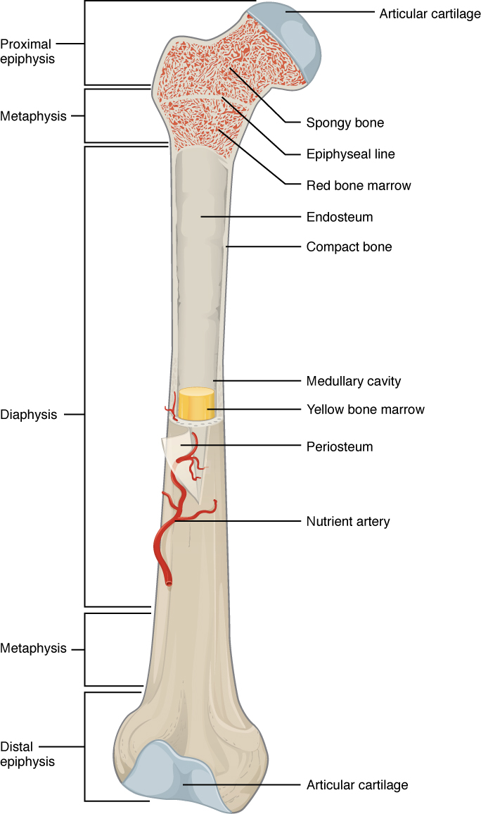
The structure of a long bone allows for the best visualization of all of the parts of a bone ( [link] ). A long bone has two parts: the diaphysis    and the epiphysis    . The diaphysis is the tubular shaft that runs between the proximal and distal ends of the bone. The hollow region in the diaphysis is called the medullary cavity    , which is filled with yellow marrow. The walls of the diaphysis are composed of dense and hard compact bone    .
Anatomy of a long bone

This illustration depicts an anterior view of the right femur, or thigh bone. The inferior end that connects to the knee is at the bottom of the diagram and the superior end that connects to the hip is at the top of the diagram. The bottom end of the bone contains a smaller lateral bulge and a larger medial bulge. A blue articular cartilage covers the inner half of each bulge as well as the small trench that runs between the bulges. This area of the inferior end of the bone is labeled the distal epiphysis. Above the distal epiphysis is the metaphysis, where the bone tapers from the wide epiphysis into the relatively thin shaft. The entire length of the shaft is the diaphysis. The superior half of the femur is cut away to show its internal contents. The bone is covered with an outer translucent sheet called the periosteum. At the midpoint of the diaphysis, a nutrient artery travels through the periosteum and into the inner layers of the bone. The periosteum surrounds a white cylinder of solid bone labeled compact bone. The cavity at the center of the compact bone is called the medullary cavity. The inner layer of the compact bone that lines the medullary cavity is called the endosteum. Within the diaphysis, the medullary cavity contains a cylinder of yellow bone marrow that is penetrated by the nutrient artery. The superior end of the femur is also connected to the diaphysis by a metaphysis. In this upper metaphysis, the bone gradually widens between the diaphysis and the proximal epiphysis. The proximal epiphysis of the femur is roughly hexagonal in shape. However, the upper right side of the hexagon has a large, protruding knob. The femur connects and rotates within the hip socket at this knob. The knob is covered with a blue colored articular cartilage. The internal anatomy of the upper metaphysis and proximal epiphysis are revealed. The medullary cavity in these regions is filled with the mesh like spongy bone. Red bone marrow occupies the many cavities within the spongy bone. There is a clear, white line separating the spongy bone of the upper metaphysis with that of the proximal epiphysis. This line is labeled the epiphyseal line.
A typical long bone shows the gross anatomical characteristics of bone.

The wider section at each end of the bone is called the epiphysis (plural = epiphyses), which is filled with spongy bone. Red marrow fills the spaces in the spongy bone. Each epiphysis meets the diaphysis at the metaphysis, the narrow area that contains the epiphyseal plate    (growth plate), a layer of hyaline (transparent) cartilage in a growing bone. When the bone stops growing in early adulthood (approximately 18–21 years), the cartilage is replaced by osseous tissue and the epiphyseal plate becomes an epiphyseal line.

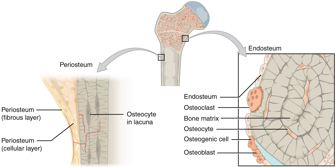
The medullary cavity has a delicate membranous lining called the endosteum    (end- = “inside”; oste- = “bone”), where bone growth, repair, and remodeling occur. The outer surface of the bone is covered with a fibrous membrane called the periosteum    (peri - = “around” or “surrounding”). The periosteum contains blood vessels, nerves, and lymphatic vessels that nourish compact bone. Tendons and ligaments also attach to bones at the periosteum. The periosteum covers the entire outer surface except where the epiphyses meet other bones to form joints ( [link] ). In this region, the epiphyses are covered with articular cartilage    , a thin layer of cartilage that reduces friction and acts as a shock absorber.
Periosteum and endosteum

The top of this illustration shows an anterior view of the proximal end of the femur. The top image has two zoom in boxes. The left box is situated on the border between the diaphysis and the metaphysis. Its callout magnifies the periosteum on the right side of the femur. The view shows that the periosteum contains an outer fibrous layer composed of yellow fibers. The inner layer of the periosteum is called the cellular layer, which is composed of irregularly shaped cells. The cellular layer gradually shrinks in width as it transitions from the metaphysis to the diaphysis. A small blood vessel runs through both layers and enters the bone. The right zoom in box magnifies the endosteum on the left side of the bone. The box is situated just inferior to the border between the diaphysis and the metaphysic. It calls out the inner edge of the compact bone layer. The magnified view shows concentric circles of dark colored bone matrix. Between the circles are small cavities containing orange, diamond-shaped cells labeled osteocytes. The left edge of the bone matrix is lined with a single layer of flattened cells called the endosteum. There is a large cell, labeled an osteoclast, between two of the endosteum cells. The osteoclast is cutting a depression into the bony matrix under the endosteum. At another part of the endosteum, three smaller osteoblasts are secreting a blue substance that builds up the outermost layer of the bony matrix.
The periosteum forms the outer surface of bone, and the endosteum lines the medullary cavity.

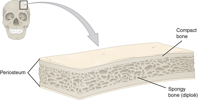
Flat bones, like those of the cranium, consist of a layer of diploë    (spongy bone), lined on either side by a layer of compact bone ( [link] ). The two layers of compact bone and the interior spongy bone work together to protect the internal organs. If the outer layer of a cranial bone fractures, the brain is still protected by the intact inner layer.
Anatomy of a flat bone

This illustration shows a cross section of a cranial bone, constructed somewhat like a sandwich. The topmost and bottommost layers are the thin, translucent, periosteum. The upper and lower periosteum cover an upper and lower layer of compact bone, respectively. The compact bone is solid, with each layer occupying about one tenth of the thickness of the cranial bone. The majority of the cross section is occupied by the spongy bone, or diploe, sandwiched between the upper and lower compact bone. The spongy bone contains many crisscrossing threads of bone. Dark air spaces occur between the threads, giving the bone a porous appearance, much like that of a sponge or Swiss cheese.
This cross-section of a flat bone shows the spongy bone (diploë) lined on either side by a layer of compact bone.

Questions & Answers

what is defense mechanism
Chinaza Reply
what is defense mechanisms
Chinaza
I'm interested in biological psychology and cognitive psychology
Tanya Reply
what does preconceived mean
sammie Reply
physiological Psychology
Nwosu Reply
How can I develope my cognitive domain
Amanyire Reply
why is communication effective
Dakolo Reply
Communication is effective because it allows individuals to share ideas, thoughts, and information with others.
effective communication can lead to improved outcomes in various settings, including personal relationships, business environments, and educational settings. By communicating effectively, individuals can negotiate effectively, solve problems collaboratively, and work towards common goals.
it starts up serve and return practice/assessments.it helps find voice talking therapy also assessments through relaxed conversation.
miss
Every time someone flushes a toilet in the apartment building, the person begins to jumb back automatically after hearing the flush, before the water temperature changes. Identify the types of learning, if it is classical conditioning identify the NS, UCS, CS and CR. If it is operant conditioning, identify the type of consequence positive reinforcement, negative reinforcement or punishment
Wekolamo Reply
please i need answer
Wekolamo
because it helps many people around the world to understand how to interact with other people and understand them well, for example at work (job).
Manix Reply
Agreed 👍 There are many parts of our brains and behaviors, we really need to get to know. Blessings for everyone and happy Sunday!
ARC
A child is a member of community not society elucidate ?
JESSY Reply
Isn't practices worldwide, be it psychology, be it science. isn't much just a false belief of control over something the mind cannot truly comprehend?
Simon Reply
compare and contrast skinner's perspective on personality development on freud
namakula Reply
Skinner skipped the whole unconscious phenomenon and rather emphasized on classical conditioning
war
explain how nature and nurture affect the development and later the productivity of an individual.
Amesalu Reply
nature is an hereditary factor while nurture is an environmental factor which constitute an individual personality. so if an individual's parent has a deviant behavior and was also brought up in an deviant environment, observation of the behavior and the inborn trait we make the individual deviant.
Samuel
I am taking this course because I am hoping that I could somehow learn more about my chosen field of interest and due to the fact that being a PsyD really ignites my passion as an individual the more I hope to learn about developing and literally explore the complexity of my critical thinking skills
Zyryn Reply
good👍
Jonathan
and having a good philosophy of the world is like a sandwich and a peanut butter 👍
Jonathan
generally amnesi how long yrs memory loss
Kelu Reply
interpersonal relationships
Abdulfatai Reply
Got questions? Join the online conversation and get instant answers!
Jobilize.com Reply

Get Jobilize Job Search Mobile App in your pocket Now!

Get it on Google Play Download on the App Store Now


Source:  OpenStax, Anatomy & Physiology: support and movement. OpenStax CNX. Aug 21, 2014 Download for free at https://legacy.cnx.org/content/col11700/1.1
Google Play and the Google Play logo are trademarks of Google Inc.

Notification Switch

Would you like to follow the 'Anatomy & Physiology: support and movement' conversation and receive update notifications?

Ask