<< Chapter < Page Chapter >> Page >
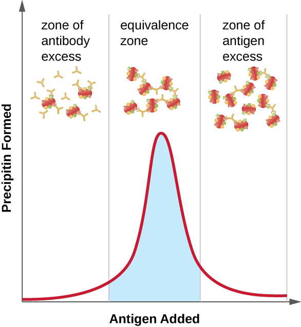
(A) Diagram of polyclonal antiserum. Antigens with multiple epitopes (shapes on their surface) are bound to different antibodies (each antibody binds to a different epitope). B) Diagram of monoclonal antibodies. Antigens with multiple epitopes have only one type of antibody bound to a single epitope on each. A graph; the X-axis is labeled antigen added and Y-axis is labeled precipitin formed. In the zone of antibody excess there is more antibody than antigen. In this case, there is no precipitate. In the equivalence zone there are approximately equal amounts of antigen and antibody. In this case a precipitate does form. In the zone of antigen excess, there is more antigen than antibody and no precipitate forms.
As antigen is slowly added to a solution containing a constant amount antibody, the amount of precipitin increases as the antibody-to-antigen ratio approaches the equivalence zone and decreases once the proportion of antigen exceeds the optimal ratio.
  • What is a precipitin?
  • Why do polyclonal antisera produce a better precipitin reaction?

Precipitin ring test

A variety of techniques allow us to use precipitin formation to quantify either antigen concentration or the amount of antibody present in an antiserum. One such technique is the precipitin ring test ( [link] ), which is used to determine the relative amount of antigen-specific antibody in a sample of serum. To perform this test, a set of test tubes is prepared by adding an antigen solution to the bottom of each tube. Each tube receives the same volume of solution, and the concentration of antigens is constant (e.g., 1 mg/mL). Next, glycerol is added to the antigen solution in each test tube, followed by a serial dilution of the antiserum. The glycerol prevents mixing of the antiserum with the antigen solution, allowing antigen-antibody binding to take place only at the interface of the two solutions. The result is a visible ring of precipitin in the tubes that have an antigen-antibody ratio within the equivalence zone. This highest dilution with a visible ring is used to determine the titer of the antibodies. The titer is the reciprocal of the highest dilution showing a positive result, expressed as a whole number. In [link] , the titer is 16.

While a measurement of titer does not tell us in absolute terms how much antibody is present, it does give a measure of biological activity, which is often more important than absolute amount. In this example, it would not be useful to know what mass of IgG were present in the antiserum, because there are many different specificities of antibody present; but it is important for us to know how much of the antibody activity in a patient’s serum is directed against the antigen of interest (e.g., a particular pathogen or allergen).
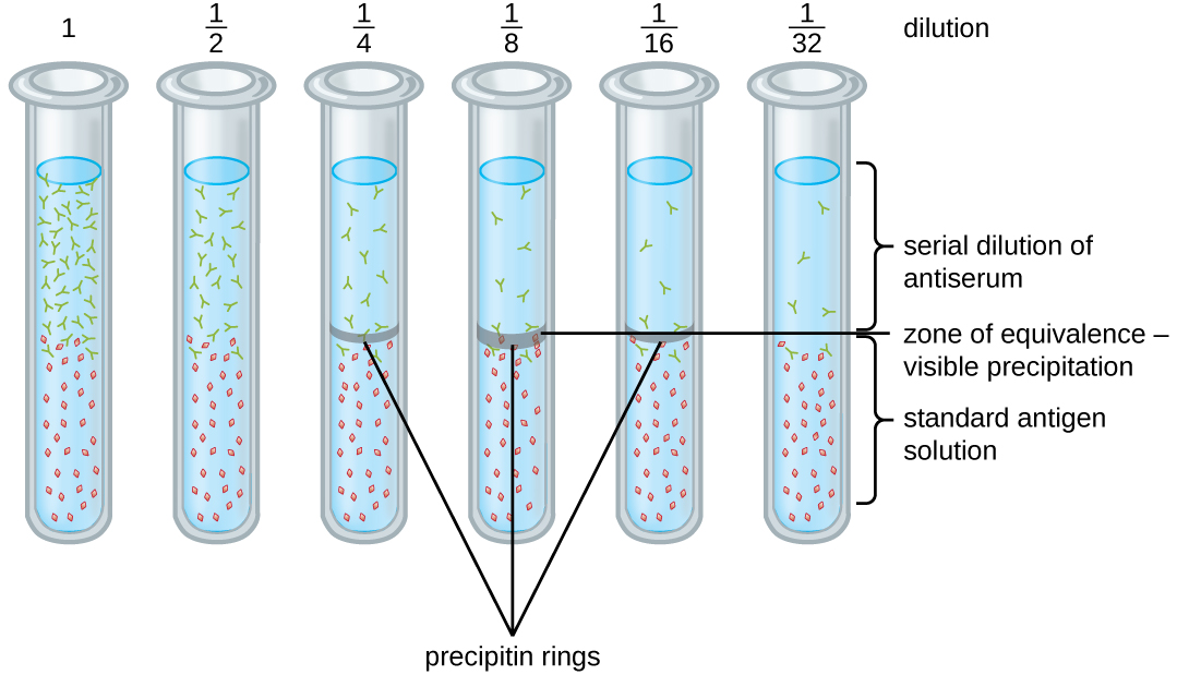
A diagram with multiple test tube dilutions labeled: 1, ½, ¼, 1/8, 1/16, 1/32. The bottom of each test tube contains a standard antigen solution. The top contains serial dilutions of antiserum. Tubes 1, ½, and 1/32 have no band at the interface between these two zones. Tubes ¼, and 1/6 have a thin band. Tube 1/8 has a thick band labeled zone of equivalence – visible precipitation.
A precipitin ring test is performed using a standard antigen solution in the bottom of the tube and a serial dilution of antiserum in the top of the tube. Glycerol prevents the two solutions from mixing so that precipitation only occurs at the interface. A visible ring of precipitation is seen in the 1/4, 1/8, and 1/16 dilutions, indicating that these concentrations are within the equivalence zone. Since 1/16 is the highest dilution in which a precipitin is observed, the titer is the reciprocal, or 16.

Ouchterlony assay

While the precipitin ring test provides insights into antibody-antigen interactions, it also has some drawbacks. It requires the use of large amounts of serum, and great care must be taken to avoid mixing the solutions and disrupting the ring. Performing a similar test in an agar gel matrix can minimize these problems. This type of assay is variously called double immunodiffusion or the Ouchterlony assay for Orjan Ouchterlony , Ouchterlony, Örjan, “In Vitro Method for Testing the Toxin-Producing Capacity of Diphtheria Bacteria,” Acta Pathologica Microbiologica Scandinavica 26, no. 4 (1949): 516-24. who first described the technique in 1948.

Questions & Answers

what are components of cells
ofosola Reply
twugzfisfjxxkvdsifgfuy7 it
Sami
58214993
Sami
what is a salt
John
the difference between male and female reproduction
John
what is computed
IBRAHIM Reply
what is biology
IBRAHIM
what is the full meaning of biology
IBRAHIM
what is biology
Jeneba
what is cell
Kuot
425844168
Sami
what is cytoplasm
Emmanuel Reply
structure of an animal cell
Arrey Reply
what happens when the eustachian tube is blocked
Puseletso Reply
what's atoms
Achol Reply
discuss how the following factors such as predation risk, competition and habitat structure influence animal's foraging behavior in essay form
Burnet Reply
cell?
Kuot
location of cervical vertebra
KENNEDY Reply
What are acid
Sheriff Reply
define biology infour way
Happiness Reply
What are types of cell
Nansoh Reply
how can I get this book
Gatyin Reply
what is lump
Chineye Reply
what is cell
Maluak Reply
what is biology
Maluak
what is vertibrate
Jeneba
what's cornea?
Majak Reply
what are cell
Achol
Got questions? Join the online conversation and get instant answers!
Jobilize.com Reply

Get Jobilize Job Search Mobile App in your pocket Now!

Get it on Google Play Download on the App Store Now


Source:  OpenStax, Microbiology. OpenStax CNX. Nov 01, 2016 Download for free at http://cnx.org/content/col12087/1.4
Google Play and the Google Play logo are trademarks of Google Inc.

Notification Switch

Would you like to follow the 'Microbiology' conversation and receive update notifications?

Ask