| << Chapter < Page | Chapter >> Page > |
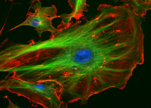
Certain materials can refract nonvisible forms of EMR and, in effect, transform them into visible light. Certain fluorescent dyes, for instance, absorb ultraviolet or blue light and then use the energy to emit photons of a different color, giving off light rather than simply vibrating. This occurs because the energy absorption causes electrons to jump to higher energy states, after which they then almost immediately fall back down to their ground states, emitting specific amounts of energy as photons. Not all of the energy is emitted in a given photon, so the emitted photons will be of lower energy and, thus, of lower frequency than the absorbed ones. Thus, a dye such as Texas red may be excited by blue light, but emit red light; or a dye such as fluorescein isothiocyanate (FITC) may absorb (invisible) high-energy ultraviolet light and emit green light ( [link] ). In some materials, the photons may be emitted following a delay after absorption; in this case, the process is called phosphorescence . Glow-in-the-dark plastic works by using phosphorescent material.

Microscopes magnify images and use the properties of light to create useful images of small objects. Magnification is defined as the ability of a lens to enlarge the image of an object when compared to the real object. For example, a magnification of 10⨯ means that the image appears 10 times the size of the object as viewed with the naked eye.
Greater magnification typically improves our ability to see details of small objects, but magnification alone is not sufficient to make the most useful images. It is often useful to enhance the resolution of objects: the ability to tell that two separate points or objects are separate. A low-resolution image appears fuzzy, whereas a high-resolution image appears sharp. Two factors affect resolution. The first is wavelength. Shorter wavelengths are able to resolve smaller objects; thus, an electron microscope has a much higher resolution than a light microscope, since it uses an electron beam with a very short wavelength, as opposed to the long-wavelength visible light used by a light microscope. The second factor that affects resolution is numerical aperture , which is a measure of a lens’s ability to gather light. The higher the numerical aperture, the better the resolution.

Read this article to learn more about factors that can increase or decrease the numerical aperture of a lens.
Even when a microscope has high resolution, it can be difficult to distinguish small structures in many specimens because microorganisms are relatively transparent. It is often necessary to increase contrast to detect different structures in a specimen. Various types of microscopes use different features of light or electrons to increase contrast—visible differences between the parts of a specimen (see Instruments of Microscopy ). Additionally, dyes that bind to some structures but not others can be used to improve the contrast between images of relatively transparent objects (see Staining Microscopic Specimens ).
When you see light bend as it moves from air into water, you are observing _________.
refraction
Explain how a prism separates white light into different colors.

Notification Switch
Would you like to follow the 'Microbiology' conversation and receive update notifications?