<< Chapter < Page Chapter >> Page >

The process by which a polypeptide chain assumes a large-scale, three-dimensional shape is called protein folding . Folded proteins that are fully functional in their normal biological role are said to possess a native structure . When a protein loses its three-dimensional shape, it may no longer be functional. These unfolded proteins are denatured . Denaturation implies the loss of the secondary structure and tertiary structure (and, if present, the quaternary structure) without the loss of the primary structure.

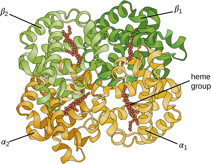
Some proteins are assemblies of several separate polypeptide s, also known as protein subunit s. These proteins function adequately only when all subunits are present and appropriately configured. The interactions that hold these subunits together constitute the quaternary structure of the protein. The overall quaternary structure is stabilized by relatively weak interactions. Hemoglobin, for example, has a quaternary structure of four globular protein subunits: two α and two β polypeptides, each one containing an iron-based heme ( [link] ).

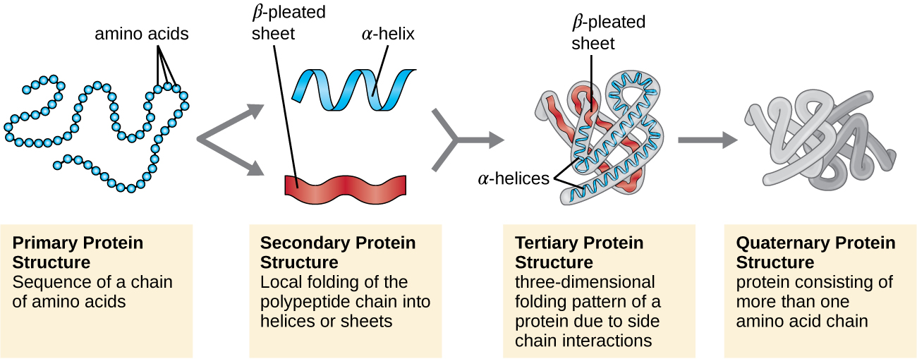
Another important class of proteins is the conjugated proteins that have a nonprotein portion. If the conjugated protein has a carbohydrate attached, it is called a glycoprotein . If it has a lipid attached, it is called a lipoprotein . These proteins are important components of membranes. [link] summarizes the four levels of protein structure.
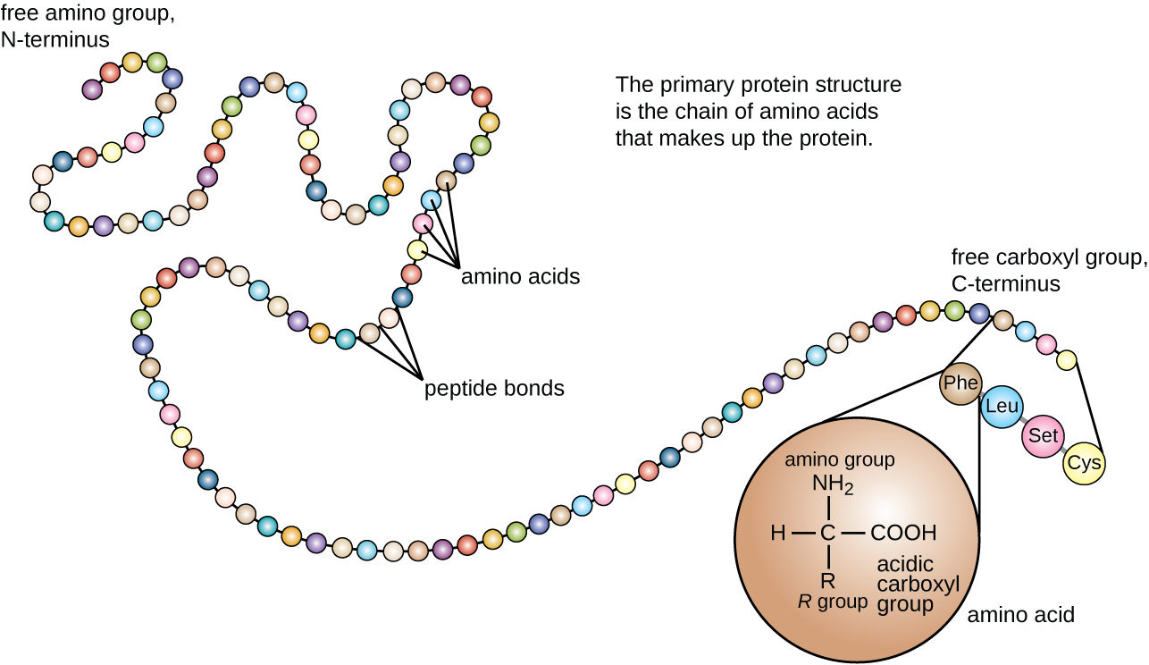
The primary protein structure is a chain of amino acids that makes up the protein. The image is a chain of circles (each circle is an amino acid). One end of the chain is the free amino group or N-terminus. The other end of the chain is the free carboxyl group or C-terminus. A drawing of a single amino acid shows a carbon with an H, an R group, a COOH (acidic carboxyl group) and an NH2 (amino group).
The primary structure of a protein is the sequence of amino acids. (credit: modification of work by National Human Genome Research Institute)
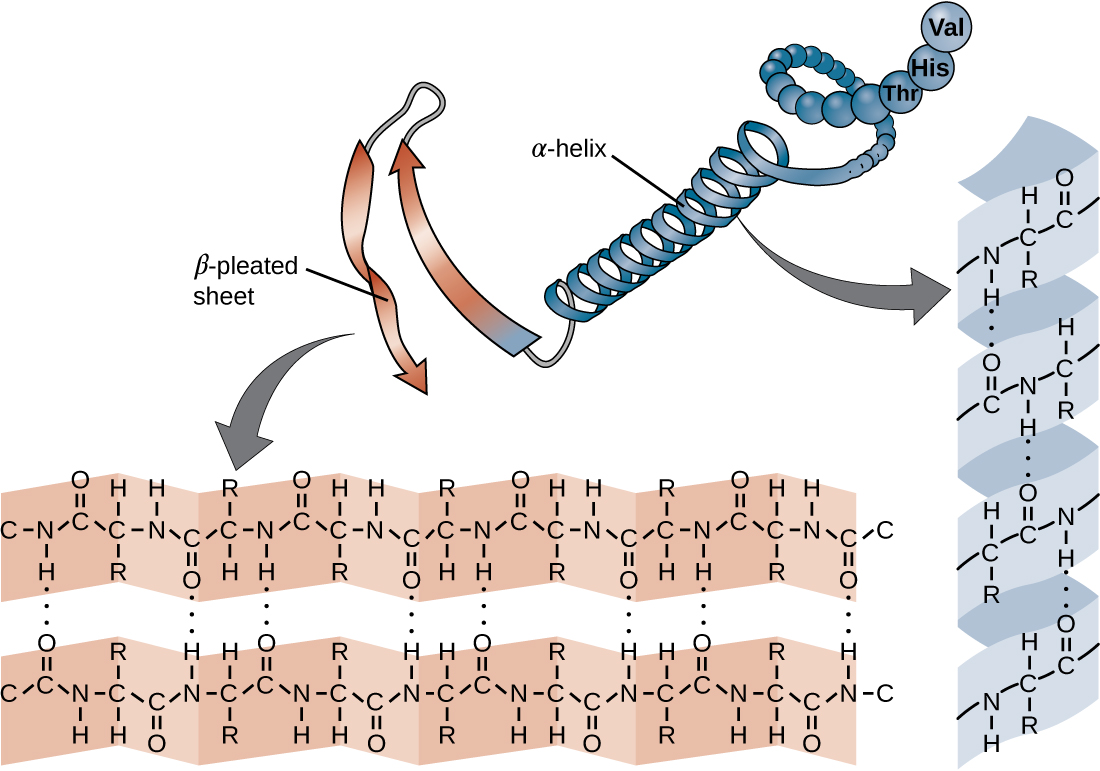
The secondary structure of a protein may be an α-helix or a β-pleated sheet, or both. A chain of spheres forms a spiral labeled alpha-helix. This chain also forms a ribbon that folds back and forth; this is labeled beta-pleated sheet. Closeups show that hydrogen bonds (dotted lines) between amino acids hold together these shapes.
The secondary structure of a protein may be an α-helix or a β-pleated sheet, or both.
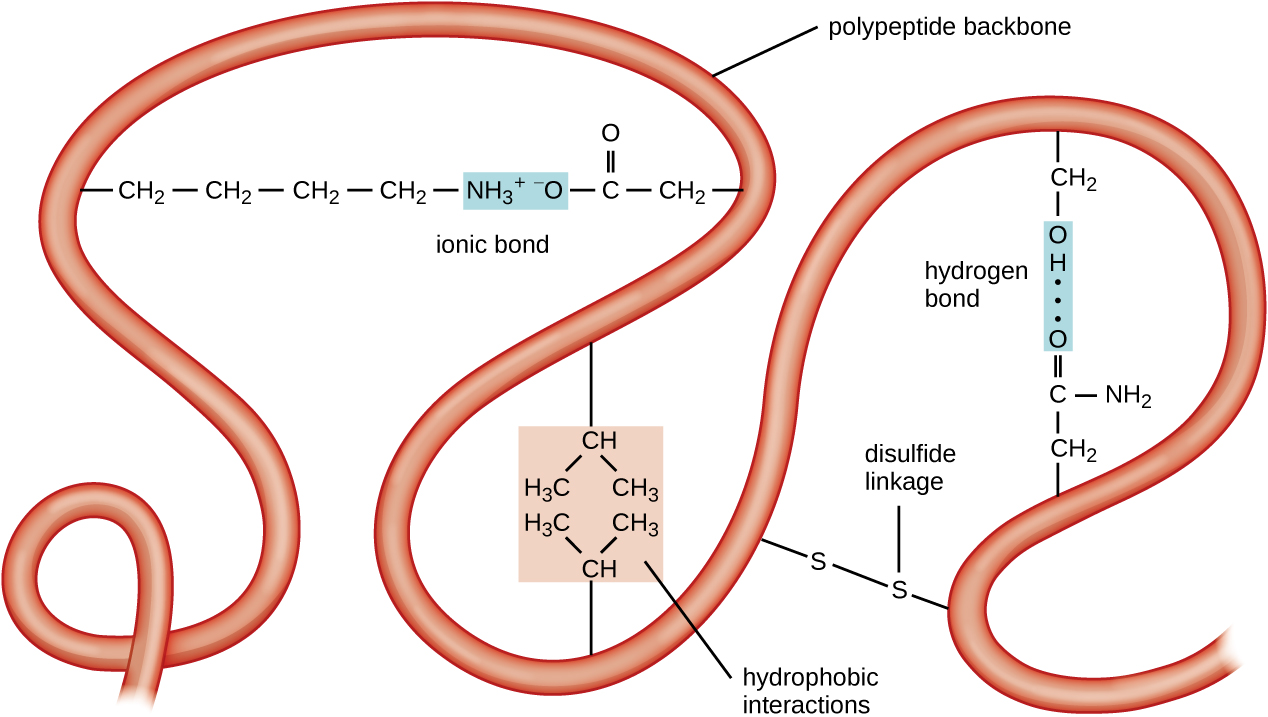
A long ribbon labeled polypeptide backbone. Loops of the ribbon are held in place by various types of chemical reactions. An ionic bond is then a positively charged amino acid and a negatively charged amino acid are attracted to each other. Hydrophobic interactions are when hydrophobic amino acids (containing only carbons and hydrogens) are clustered together. A disulfide linkage is when a sulfur of one amino acid is covalently bound to the sulfur of another amino acid. A hydrogen bond is when two polar amino acids are attracted to each other.
The tertiary structure of proteins is determined by a variety of attractive forces, including hydrophobic interactions, ionic bonding, hydrogen bonding, and disulfide linkages.
A complex spherical shape made of ribbons that are coiled and wound around each other. There are 4 large regions (each made from a separate ribbon) – alpha 1, alpha 2, beta 1, beta 2.  There are also red spheres attached to each ribbon; these are labeled heme group.
A hemoglobin molecule has two α and two β polypeptides together with four heme groups.
Primary protein structure: sequence of a chain of amino acids. This is shown as a chain of circles. Secondary protein structure: local folding of the polypeptide chain into helices or sheets. This is shown as a spiral labeled alpha-helix and a folded sheet labeled beta-pleated sheet. Tertiary protein structure: three-dimensional folding pattern of a protein due to side chain interactions. This is shown as a complex 3-D shape made of alpha helices and beta pleated sheets. Quaternary protein structure: protein consisting of more than one amino acid chain. This is shown as 2 complex structures similar to that seen at the tertiary level.
Protein structure has four levels of organization. (credit: modification of work by National Human Genome Research Institute)
  • What can happen if a protein’s primary, secondary, tertiary, or quaternary structure is changed?

Primary structure, dysfunctional proteins, and cystic fibrosis

Proteins associated with biological membranes are classified as extrinsic or intrinsic. Extrinsic proteins, also called peripheral proteins, are loosely associated with one side of the membrane. Intrinsic proteins, or integral proteins, are embedded in the membrane and often function as part of transport systems as transmembrane proteins. Cystic fibrosis (CF) is a human genetic disorder caused by a change in the transmembrane protein. It affects mostly the lungs but may also affect the pancreas, liver, kidneys, and intestine. CF is caused by a loss of the amino acid phenylalanine in a cystic fibrosis transmembrane protein (CFTR). The loss of one amino acid changes the primary structure of a protein that normally helps transport salt and water in and out of cells ( [link] ).

The change in the primary structure prevents the protein from functioning properly, which causes the body to produce unusually thick mucus that clogs the lungs and leads to the accumulation of sticky mucus. The mucus obstructs the pancreas and stops natural enzymes from helping the body break down food and absorb vital nutrients.

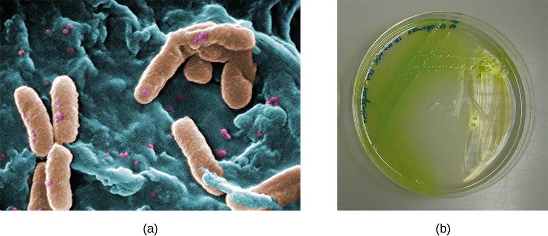
In the lungs of individuals with cystic fibrosis, the altered mucus provides an environment where bacteria can thrive. This colonization leads to the formation of biofilms in the small airways of the lungs. The most common pathogens found in the lungs of patients with cystic fibrosis are Pseudomonas aeruginosa ( [link] ) and Burkholderia cepacia . Pseudomonas differentiates within the biofilm in the lung and forms large colonies, called “mucoid” Pseudomonas . The colonies have a unique pigmentation that shows up in laboratory tests ( [link] ) and provides physicians with the first clue that the patient has CF (such colonies are rare in healthy individuals).
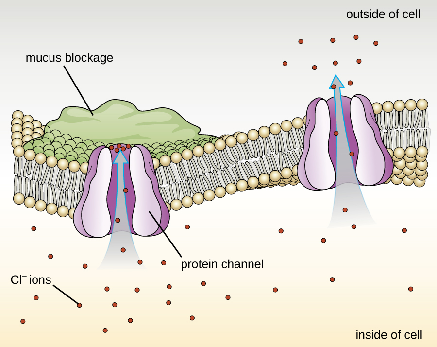
A drawing of a phospholipid bilayer in the center with two protein channels. One is open and lets Cl- flow out of the cell. The other is blocked by a mucus blockage on the outside of the cell; Cl- ions can’t flow through this channel.
The normal CFTR protein is a channel protein that helps salt (sodium chloride) move in and out of cells.
a) a micrograph of rod shaped cells. B) An agar plate with a green pigmented colonies; this green pigment is spreading past the edge of the colonies.
(a) A scanning electron micrograph shows the opportunistic bacterium Pseudomonas aeruginosa . (b) Pigment-producing P. aeruginosa on cetrimide agar shows the green pigment called pyocyanin. (credit a: modification of work by the Centers for Disease Control and Prevention)

Key concepts and summary

  • Amino acids are small molecules essential to all life. Each has an α carbon to which a hydrogen atom, carboxyl group, and amine group are bonded. The fourth bonded group, represented by R, varies in chemical composition, size, polarity, and charge among different amino acids, providing variation in properties.
  • Peptides are polymers formed by the linkage of amino acids via dehydration synthesis. The bonds between the linked amino acids are called peptide bonds. The number of amino acids linked together may vary from a few to many.
  • Proteins are polymers formed by the linkage of a very large number of amino acids. They perform many important functions in a cell, serving as nutrients and enzymes; storage molecules for carbon, nitrogen, and energy; and structural components.
  • The structure of a protein is a critical determinant of its function and is described by a graduated classification: primary , secondary , tertiary , and quaternary . The native structure of a protein may be disrupted by denaturation , resulting in loss of its higher-order structure and its biological function.
  • Some proteins are formed by several separate protein subunits, the interaction of these subunits composing the quaternary structure of the protein complex.
  • Conjugated proteins have a nonpolypeptide portion that can be a carbohydrate (forming a glycoprotein ) or a lipid fraction (forming a lipoprotein ). These proteins are important components of membranes.

Fill in the blank

The sequence of amino acids in a protein is called its __________.

Primary structure

Got questions? Get instant answers now!

Denaturation implies the loss of the __________ and __________ structures without the loss of the __________ structure.

secondary, tertiary, primary

Got questions? Get instant answers now!

True/false

A change in one amino acid in a protein sequence always results in a loss of function.

False

Got questions? Get instant answers now!

Get Jobilize Job Search Mobile App in your pocket Now!

Get it on Google Play Download on the App Store Now


Source:  OpenStax, Microbiology. OpenStax CNX. Nov 01, 2016 Download for free at http://cnx.org/content/col12087/1.4
Google Play and the Google Play logo are trademarks of Google Inc.

Notification Switch

Would you like to follow the 'Microbiology' conversation and receive update notifications?

Ask