<< Chapter < Page Chapter >> Page >

Learning objectives

  • Identify and describe the components of blood
  • Explain the process by which the formed elements of blood are formed (hematopoiesis)
  • Describe the characteristics of formed elements found in peripheral blood, as well as their respective functions within the innate immune system

In the previous section, we discussed some of the chemical mediators found in plasma, the fluid portion of blood. The nonfluid portion of blood consists of various types of formed elements , so called because they are all formed from the same stem cells found in bone marrow. The three major categories of formed elements are: red blood cells (RBCs), also called erythrocyte s ; platelets , also called thrombocytes ; and white blood cells (WBCs), also called leukocytes .

Red blood cells are primarily responsible for carrying oxygen to tissues. Platelets are cellular fragments that participate in blood clot formation and tissue repair. Several different types of WBCs participate in various nonspecific mechanisms of innate and adaptive immunity. In this section, we will focus primarily on the innate mechanisms of various types of WBCs.

Hematopoiesis

All of the formed elements of blood are derived from pluripotent hematopoietic stem cells (HSCs) in the bone marrow. As the HSCs make copies of themselves in the bone marrow, individual cells receive different cues from the body that control how they develop and mature. As a result, the HSCs differentiate into different types of blood cells that, once mature, circulate in peripheral blood. This process of differentiation, called hematopoiesis , is shown in more detail in [link] .

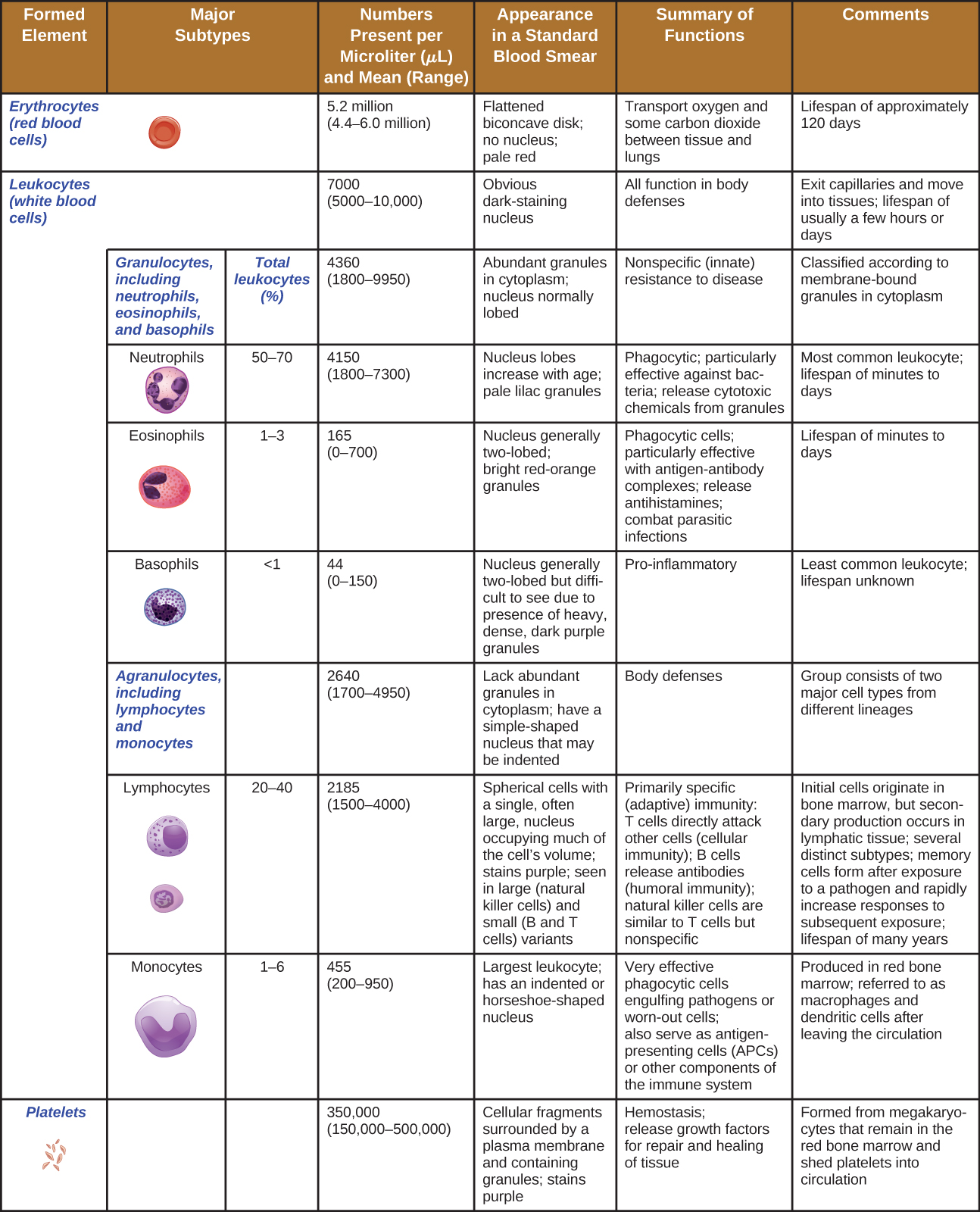
In terms of sheer numbers, the vast majority of HSCs become erythrocytes. Much smaller numbers become leukocytes and platelets. Leukocytes can be further subdivided into granulocytes , which are characterized by numerous granules visible in the cytoplasm, and agranulocytes , which lack granules. [link] provides an overview of the various types of formed elements, including their relative numbers, primary function, and lifespans.
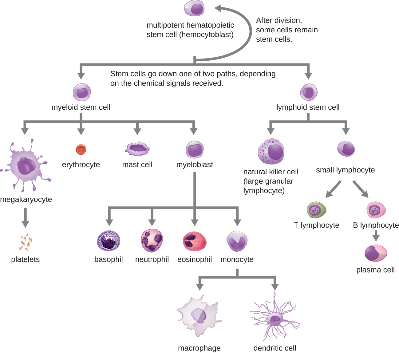
A flowchart showing progression of development for formed elements of blood. At the top is a multipotent hematopoietic stem cell (hemocytoblast). This cell divides and after division some of the new cells remain stem cells. Others go down one of two paths depending on the chemical signals received. One path begins with lymphoid stem cells which can either become natural killer cells (large granular lymphocytes) or small lymphocytes. The natural killer cell is a medium-large purple cell. Small lymphocytes can either become T lymphocytes or B lymphoctyes. The T and B lymphocytes are medium size cells with a large nucleus. B lymphocytes become plasma cells which are medium size cells with a large nucleus. The other option for the stem cell is to become a myeloid stem cell. Myeloid stem cells follow one of four paths. One path leads to megakaryocyte which leads to platelets. Platelets are small flecks. The second path leads to erythrocyte. Erythrocytes are small donut shaped red cells. The third path leads to mast cells. The fourth path leads to basophil, neutrophil, eosinophil, or monocyte. Basophils are medium cells with many dark purple spots. Neutrophils are medium pink cells with a multi-lobbed nucleus. Eosinophils are medium size cells with many pink spots. Monocytes lead to macrophages or dendritic cells. Macrophages are large irregularly shaped cells. Dendritic cells have longer tendons branching off of them.
All the formed elements of the blood arise by differentiation of hematopoietic stem cells in the bone marrow.
A table of the formed elements. Top row reads: formed element, major subtypes, numbers present per microliter and mean, appearance in standard blood smear, summary of functions, and comments. The first row is for erythrocytes (red blood cells). There are 5.2 million per microliter of blood (ranging from 4.4 – 6 million). These cells are flattened biconcave disks with no nucleus and a pale red color. Their function is to transport oxygen and some carbon dioxide between tissue and lungs. Their lifespan is approximately 120 days. The set of rows is classified under leukocytes (white blood cells). Leukocytes as a group number 7000 per microliter of blood (ranging from 5000-10,000). Leukocytes have an obvious dark-staining nucleus and function in body defenses. They exit capillaries and move into tissues. Their lifespan is usually a few hours or days. Leukocytes are divided into two groups. The first is granulocytes including neutrophils, eosinophils, and basophils. The second is agranulocytes including lymphocytes and monocytes. Granulocytes number 4300 per microliter of blood (range of 1800-9950). Granulocytes have abundant granules in the cytoplasm and the nucleus is normally lobed. Granulocytes function in nonspecific (innate) resistance to disease and are classified according to membrane-bound granules in the cytoplasm. Neutrophils make up 50-70% of the total leukocytes and number 4150 per microliter of blood (range 1800-7300). Neutrophils have a nucleus with lobes that increase with age and pale lilac granules. They are phagocytic and particularly effective against bacteria; they release toxic chemicals from granules. Neutrophils are the most common leukocyte with a lifespan of minutes to days. Eosinophils make up 1-3% of total leukocytes. They number 165 per microliter of blood (range of 0 – 700). Eosinophils have a nucleus that is generally two-lobed and bright red-orange granules. They are phagocytic cells and particularly effective with antigen-antibody complexes. Eosinophils release antihistamines and increase allergies, they also help fight parasitic infections. Eosinophils have a lifespan of minutes to days. Basophils make up less than 1% of total leukocytes. They number 44 per microliter of blood (range 0 – 150). Basophils have a nucleus that is generally two lobed but difficult to see due to the presence of heavy, dense, dark purple granules. Basophils promote inflammation and are the least common leukocyte. Their lifespan is unknown. Agranulocytes (including lymphocytes and monocytes) number 2640 per microliter of blood (range 1700 – 4900). Agranulocytes lack abundant granules in the cytoplasm and have a simple-shaped nucleus that may be indented. They function in body defenses and are grouped into two major cell types from different lineages. Lymphocytes make up 20-40% of total leukocytes and number 2185 per microliter of blood (range 1500-4000). Lymphocytes are spherical cells with a single, often large, nucleus occupying much of the cell’s volume. They stain purple and are seen in large (natural killer cells) and small (B and T cells) variants. Lymphocytes are primarily involved in specific (adaptive) immunity. T cells directly attack other cells (cellular immunity); natural killer cells are similar to T cells but nonspecific. Lymphocytes originate in bone marrow but secondary production occurs in lymphatic tissue. Several distinct subtypes. Memory cells form after exposure to a pathogen and rapidly increase responses to subsequent exposure. Lifespan of many years. Monocytes make up 1-6% of total leukocytes. They number 455 per microliter of blood (range 200-950). Monocytes are large leukocytes with an indented or horseshoe-shaped nucleus. They are very effective phagocytic cells engulfing pathogens or worn out cells and also serve as antigen presenting cells (APCs) or other components of the immune system. Monocytes are produced in red bone marrow and are referred to as macrophages and dendritic cells after leaving circulation. The last row of the table is for platelets. These number 350,000 per microliter of blood (range 150,000-500,000). Platelets are cellular fragments surrounded by a plasma membrane and containing granules. They stain purple. The function of platelets is hemostasis plus releasing growth factors for repair and healing of tissues. They are formed from megakaryocytes that remain in the red bone marrow and shed platelets into circulation.
Formed elements of blood include erythrocytes (red blood cells), leukocytes (white blood cells), and platelets.

Granulocytes

The various types of granulocytes can be distinguished from one another in a blood smear by the appearance of their nuclei and the contents of their granules, which confer different traits, functions, and staining properties. The neutrophils , also called polymorphonuclear neutrophils (PMNs) , have a nucleus with three to five lobes and small, numerous, lilac-colored granules. Each lobe of the nucleus is connected by a thin strand of material to the other lobes. The eosinophils have fewer lobes in the nucleus (typically 2–3) and larger granules that stain reddish-orange. The basophils have a two-lobed nucleus and large granules that stain dark blue or purple ( [link] ).

Neutrophils have a multi-lobed nucleus. Eosinophils have a two-lobed nucleus and distinct pink spots when stained. Basophils have a two-lobed nucleus and distinct purple spots when stained. Each type of granulocyte is illustrated with a micrograph above it.
Granulocytes can be distinguished by the number of lobes in their nuclei and the staining properties of their granules. (credit “neutrophil” micrograph: modification of work by Ed Uthman)

Get Jobilize Job Search Mobile App in your pocket Now!

Get it on Google Play Download on the App Store Now


Source:  OpenStax, Microbiology. OpenStax CNX. Nov 01, 2016 Download for free at http://cnx.org/content/col12087/1.4
Google Play and the Google Play logo are trademarks of Google Inc.

Notification Switch

Would you like to follow the 'Microbiology' conversation and receive update notifications?

Ask