<< Chapter < Page Chapter >> Page >

Bone marrow

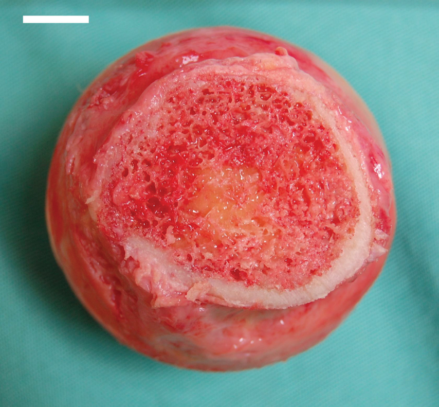
This photograph shows the bone marrow.
Red bone marrow fills the head of the femur, and a spot of yellow bone marrow is visible in the center. The white reference bar is 1 cm.

Thymus

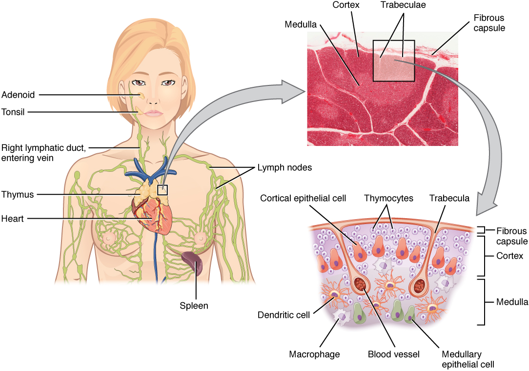
The thymus    gland is a bilobed organ found in the space between the sternum and the aorta of the heart ( [link] ). Connective tissue holds the lobes closely together but also separates them and forms a capsule.
Location, structure, and histology of the thymus

The left panel of this figure shows the head and chest of a woman and the location of the thymus is marked. The top right panel shows a micrograph of the thymus and the bottom right panel shows a magnified view of the structure of the thymus.
The thymus lies above the heart. The trabeculae and lobules, including the darkly staining cortex and the lighter staining medulla of each lobule, are clearly visible in the light micrograph of the thymus of a newborn. LM × 100. (Micrograph provided by the Regents of the University of Michigan Medical School © 2012)

View the University of Michigan WebScope at (External Link) to explore the tissue sample in greater detail.

The connective tissue capsule further divides the thymus into lobules via extensions called trabeculae. The outer region of the organ is known as the cortex and contains large numbers of thymocytes with some epithelial cells, macrophages, and dendritic cells (two types of phagocytic cells that are derived from monocytes). The cortex is densely packed so it stains more intensely than the rest of the thymus (see [link] ). The medulla, where thymocytes migrate before leaving the thymus, contains a less dense collection of thymocytes, epithelial cells, and dendritic cells.

Aging and the…

Immune system

By the year 2050, 25 percent of the population of the United States will be 60 years of age or older. The CDC estimates that 80 percent of those 60 years and older have one or more chronic disease associated with deficiencies of the immune systems. This loss of immune function with age is called immunosenescence. To treat this growing population, medical professionals must better understand the aging process. One major cause of age-related immune deficiencies is thymic involution, the shrinking of the thymus gland that begins at birth, at a rate of about three percent tissue loss per year, and continues until 35–45 years of age, when the rate declines to about one percent loss per year for the rest of one’s life. At that pace, the total loss of thymic epithelial tissue and thymocytes would occur at about 120 years of age. Thus, this age is a theoretical limit to a healthy human lifespan.

Thymic involution has been observed in all vertebrate species that have a thymus gland. Animal studies have shown that transplanted thymic grafts between inbred strains of mice involuted according to the age of the donor and not of the recipient, implying the process is genetically programmed. There is evidence that the thymic microenvironment, so vital to the development of naïve T cells, loses thymic epithelial cells according to the decreasing expression of the FOXN1 gene with age.

It is also known that thymic involution can be altered by hormone levels. Sex hormones such as estrogen and testosterone enhance involution, and the hormonal changes in pregnant women cause a temporary thymic involution that reverses itself, when the size of the thymus and its hormone levels return to normal, usually after lactation ceases. What does all this tell us? Can we reverse immunosenescence, or at least slow it down? The potential is there for using thymic transplants from younger donors to keep thymic output of naïve T cells high. Gene therapies that target gene expression are also seen as future possibilities. The more we learn through immunosenescence research, the more opportunities there will be to develop therapies, even though these therapies will likely take decades to develop. The ultimate goal is for everyone to live and be healthy longer, but there may be limits to immortality imposed by our genes and hormones.

Questions & Answers

what are the types of homeostasis
Odey Reply
diagram of the digestive system
Zainab Reply
drown and level female reproductive system
Anas
anatomy
Anas
What is the best way to indicate the sperm
ADAM Reply
Definition of pathology
Promise Reply
what are the body organs and their functions
Comforter Reply
what are the body organs and their functions
Ruth
musculoskeletal
Ruth
what is cell
Oppicial Reply
a cell is the smallest structural and functional unit of life.
Patrick
To know how bones are functions
DAUDA Reply
diagram of the heart
Victoria Reply
what are the layers of the muscles
Tongdock Reply
What is Amebae
Najibu Reply
the collection of fluids in the throat is cause by what
Emmanuel Reply
what is difference between meiosis and mitosis
Aishetu Reply
what is difference between mitosis and meiosis
Aishetu
What is Anatomy
Najibu Reply
What the difference between the Anatomy and physiology
Najibu
What is the meaning of chromoprotein
Aisha Reply
what is cartilage
Abdulkadir Reply
tough , white fibrous tissue
Henry
drowning and level female reproductive system
Anas Reply

Get Jobilize Job Search Mobile App in your pocket Now!

Get it on Google Play Download on the App Store Now


Source:  OpenStax, Anatomy & Physiology. OpenStax CNX. Feb 04, 2016 Download for free at http://legacy.cnx.org/content/col11496/1.8
Google Play and the Google Play logo are trademarks of Google Inc.

Notification Switch

Would you like to follow the 'Anatomy & Physiology' conversation and receive update notifications?

Ask