| << Chapter < Page | Chapter >> Page > |

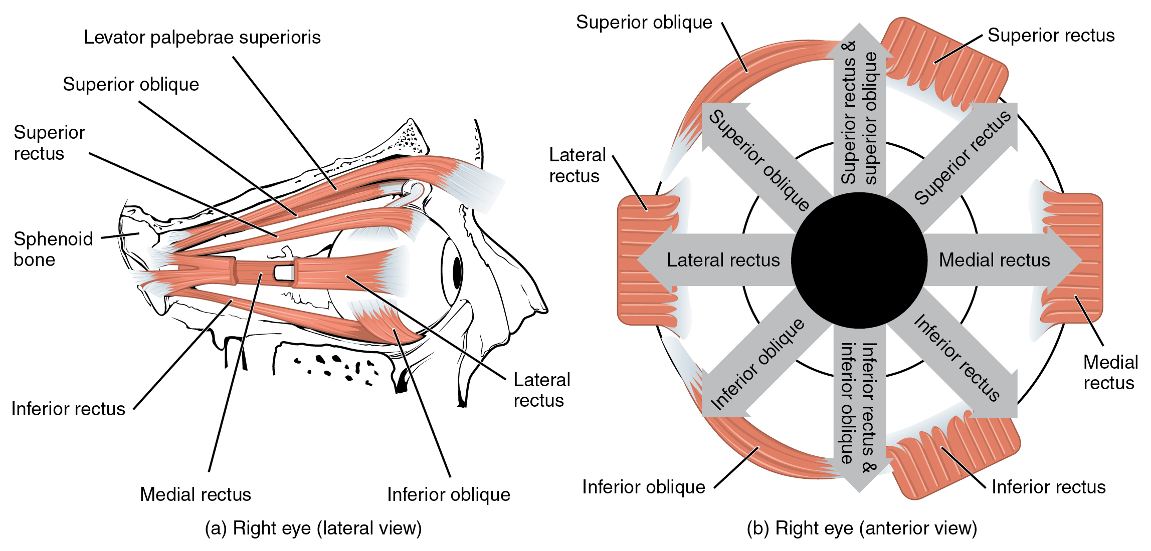
| Muscles of the Eyes | |||||
|---|---|---|---|---|---|
| Movement | Target | Target motion direction | Prime mover | Origin | Insertion |
| Moves eyes up and toward nose; rotates eyes from 1 o’clock to 3 o’clock | Eyeballs | Superior (elevates); medial (adducts) | Superior rectus | Common tendinous ring (ring attaches to optic foramen) | Superior surface of eyeball |
| Moves eyes down and toward nose; rotates eyes from 6 o’clock to 3 o’clock | Eyeballs | Inferior (depresses); medial (adducts) | Inferior rectus | Common tendinous ring (ring attaches to optic foramen) | Inferior surface of eyeball |
| Moves eyes away from nose | Eyeballs | Lateral (abducts) | Lateral rectus | Common tendinous ring (ring attaches to optic foramen) | Lateral surface of eyeball |
| Moves eyes toward nose | Eyeballs | Medial (adducts) | Medial rectus | Common tendinous ring (ring attaches to optic foramen) | Medial surface of eyeball |
| Moves eyes up and away from nose; rotates eyeball from 12 o’clock to 9 o’clock | Eyeballs | Superior (elevates); lateral (abducts) | Inferior oblique | Floor of orbit (maxilla) | Surface of eyeball between inferior rectus and lateral rectus |
| Moves eyes down and away from nose; rotates eyeball from 6 o’clock to 9 o’clock | Eyeballs | Superior (elevates); lateral (abducts) | Superior oblique | Sphenoid bone | Suface of eyeball between superior rectus and lateral rectus |
| Opens eyes | Upper eyelid | Superior (elevates) | Levator palpabrae superioris | Roof of orbit (sphenoid bone) | Skin of upper eyelids |
| Closes eyelids | Eyelid skin | Compression along superior–inferior axis | Orbicularis oculi | Medial bones composing the orbit | Circumference of orbit |
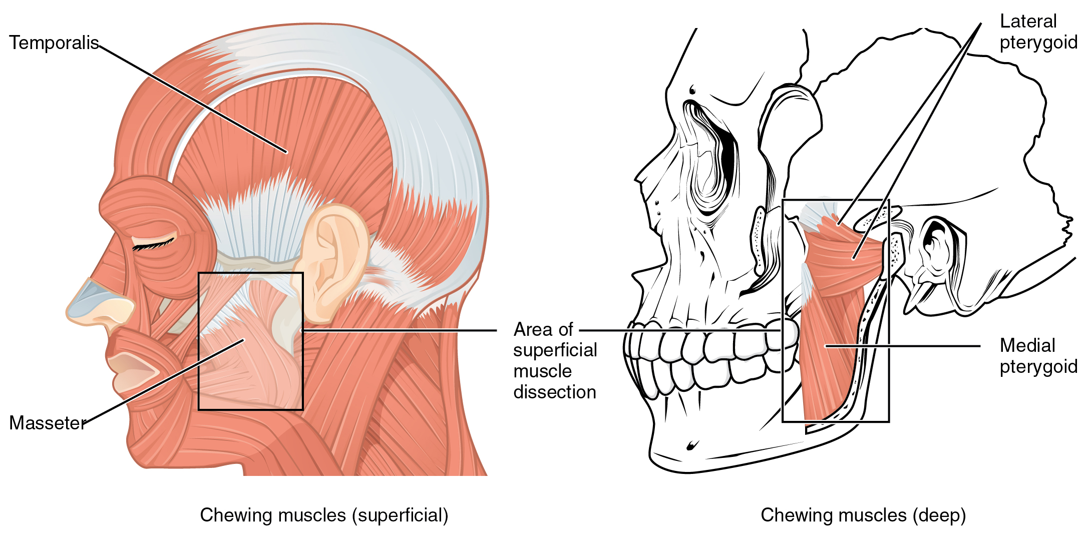
In anatomical terminology, chewing is called mastication . Muscles involved in chewing must be able to exert enough pressure to bite through and then chew food before it is swallowed ( [link] and [link] ). The masseter muscle is the main muscle used for chewing because it elevates the mandible (lower jaw) to close the mouth, and it is assisted by the temporalis muscle, which retracts the mandible. You can feel the temporalis move by putting your fingers to your temple as you chew.

| Muscles of the Lower Jaw | |||||
|---|---|---|---|---|---|
| Movement | Target | Target motion direction | Prime mover | Origin | Insertion |
| Closes mouth; aids chewing | Mandible | Superior (elevates) | Masseter | Maxilla arch; zygomatic arch (for masseter) | Mandible |
| Closes mouth; pulls lower jaw in under upper jaw | Mandible | Superior (elevates); posterior (retracts) | Temporalis | Temporal bone | Mandible |
| Opens mouth; pushes lower jaw out under upper jaw; moves lower jaw side-to-side | Mandible | Inferior (depresses); posterior (protracts); lateral (abducts); medial (adducts) | Lateral pterygoid | Pterygoid process of sphenoid bone | Mandible |
| Closes mouth; pushes lower jaw out under upper jaw; moves lower jaw side-to-side | Mandible | Superior (elevates); posterior (protracts); lateral (abducts); medial (adducts) | Medial pterygoid | Sphenoid bone; maxilla | Mandible; temporo-mandibular joint |

Notification Switch
Would you like to follow the 'Anatomy & Physiology' conversation and receive update notifications?