<< Chapter < Page Chapter >> Page >
By the end of this section, you will be able to:
  • Describe the structures of DNA and RNA
  • Describe how eukaryotic DNA is arranged in the cell

In the 1950s, Francis Crick and James Watson worked together at the University of Cambridge, England, to determine the structure of DNA. Other scientists, such as Linus Pauling and Maurice Wilkins, were also actively exploring this field. Pauling had discovered the secondary structure of proteins using X-ray crystallography. X-ray crystallography is a method for investigating molecular structure by observing the patterns formed by X-rays shot through a crystal of the substance. The patterns give important information about the structure of the molecule of interest. In Wilkins’ lab, researcher Rosalind Franklin was using X-ray crystallography to understand the structure of DNA. Watson and Crick were able to piece together the puzzle of the DNA molecule using Franklin's data ( [link] ). Watson and Crick also had key pieces of information available from other researchers such as Chargaff’s rules. Chargaff had shown that of the four kinds of monomers (nucleotides) present in a DNA molecule, two types were always present in equal amounts and the remaining two types were also always present in equal amounts. This meant they were always paired in some way. In 1962, James Watson, Francis Crick, and Maurice Wilkins were awarded the Nobel Prize in Medicine for their work in determining the structure of DNA.
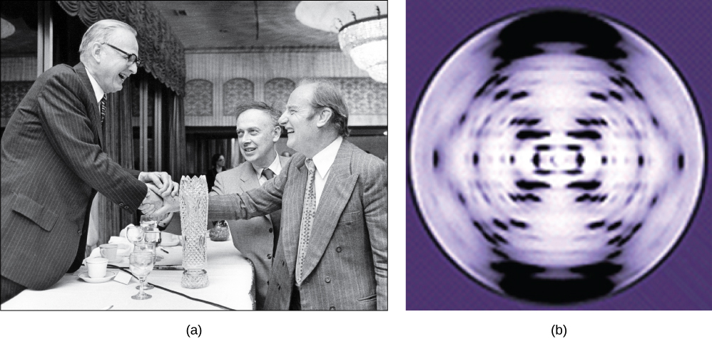
Photo in part A shows James Watson, Francis Crick, and Maclyn McCarty. The x-ray diffraction pattern in part b is symmetrical, with dots in an x-shape.
Pioneering scientists (a) James Watson and Francis Crick are pictured here with American geneticist Maclyn McCarty. Scientist Rosalind Franklin discovered (b) the X-ray diffraction pattern of DNA, which helped to elucidate its double helix structure. (credit a: modification of work by Marjorie McCarty; b: modification of work by NIH)

Now let’s consider the structure of the two types of nucleic acids, deoxyribonucleic acid (DNA) and ribonucleic acid (RNA). The building blocks of DNA are nucleotides, which are made up of three parts: a deoxyribose    (a 5-carbon sugar with the carbons designated as 1', 2', 3', 4' and 5'), a phosphate group    attached to the 5'carbon, and a nitrogenous base    attached to the 1'carbon( [link] ). There are four types of nitrogenous bases in DNA. Adenine (A) and guanine (G) are double-ringed purines, and cytosine (C) and thymine (T) are smaller, single-ringed pyrimidines. The nucleotide is named according to the nitrogenous base it contains.

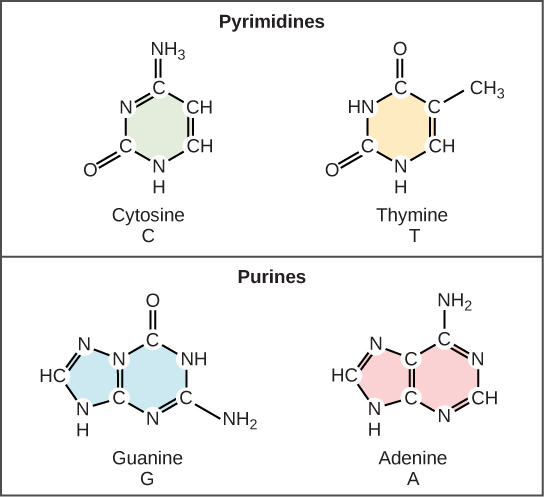
Illustration shows structure of a nucleotide, which is made up of a deoxyribose sugar with a nitrogenous base attached at the 1' position and a phosphate group attached at the 5' position. There are two kinds of nitrogenous bases: pyrimidines, which have one six-membered ring, and purines, which have a six-membered ring fused to a five-membered ring. Cytosine and thymine are pyrimidines, and adenine and guanine are purines.
(a) Each DNA nucleotide is made up of a sugar, a phosphate group, and a base. Four of the five carbon atoms in the sugar are not shown to simplify the diagram; each point on the ring is where a carbon (1' to 4') would be would be located; the position of the 5' carbon is above the ring at the point adjacent to an oxygen of the phosphate group. (b) Cytosine and thymine are pyrimidines. Guanine and adenine are purines. Note: As the 2' carbon is attached to an -OH group, the sugar shown is actually ribose, not deoxyribose as intended.

The phosphate group of one nucleotide bonds covalently with the sugar molecule of the next nucleotide, and so on, forming a long polymer of nucleotide monomers. The sugar–phosphate groups line up in a “backbone” for each single strand of DNA, and the nucleotide bases stick out from this backbone. The carbon atoms of the five-carbon sugar are numbered clockwise from the oxygen as 1', 2', 3', 4', and 5' (1' is read as “one prime”). The phosphate group is attached to the 5' carbon of one nucleotide and the 3' carbon of the next nucleotide. In its natural state, each DNA molecule is actually composed of two single strands held together along their length with hydrogen bonds between the bases.

Questions & Answers

what are components of cells
ofosola Reply
twugzfisfjxxkvdsifgfuy7 it
Sami
58214993
Sami
what is a salt
John
the difference between male and female reproduction
John
what is computed
IBRAHIM Reply
what is biology
IBRAHIM
what is the full meaning of biology
IBRAHIM
what is biology
Jeneba
what is cell
Kuot
425844168
Sami
what is biology
Inenevwo
what is cytoplasm
Emmanuel Reply
structure of an animal cell
Arrey Reply
what happens when the eustachian tube is blocked
Puseletso Reply
what's atoms
Achol Reply
discuss how the following factors such as predation risk, competition and habitat structure influence animal's foraging behavior in essay form
Burnet Reply
cell?
Kuot
location of cervical vertebra
KENNEDY Reply
What are acid
Sheriff Reply
define biology infour way
Happiness Reply
What are types of cell
Nansoh Reply
how can I get this book
Gatyin Reply
what is lump
Chineye Reply
what is cell
Maluak Reply
what is biology
Maluak
what is vertibrate
Jeneba
what's cornea?
Majak Reply
what are cell
Achol
Got questions? Join the online conversation and get instant answers!
Jobilize.com Reply

Get Jobilize Job Search Mobile App in your pocket Now!

Get it on Google Play Download on the App Store Now


Source:  OpenStax, Human biology. OpenStax CNX. Dec 01, 2015 Download for free at http://legacy.cnx.org/content/col11903/1.3
Google Play and the Google Play logo are trademarks of Google Inc.

Notification Switch

Would you like to follow the 'Human biology' conversation and receive update notifications?

Ask