| << Chapter < Page | Chapter >> Page > |
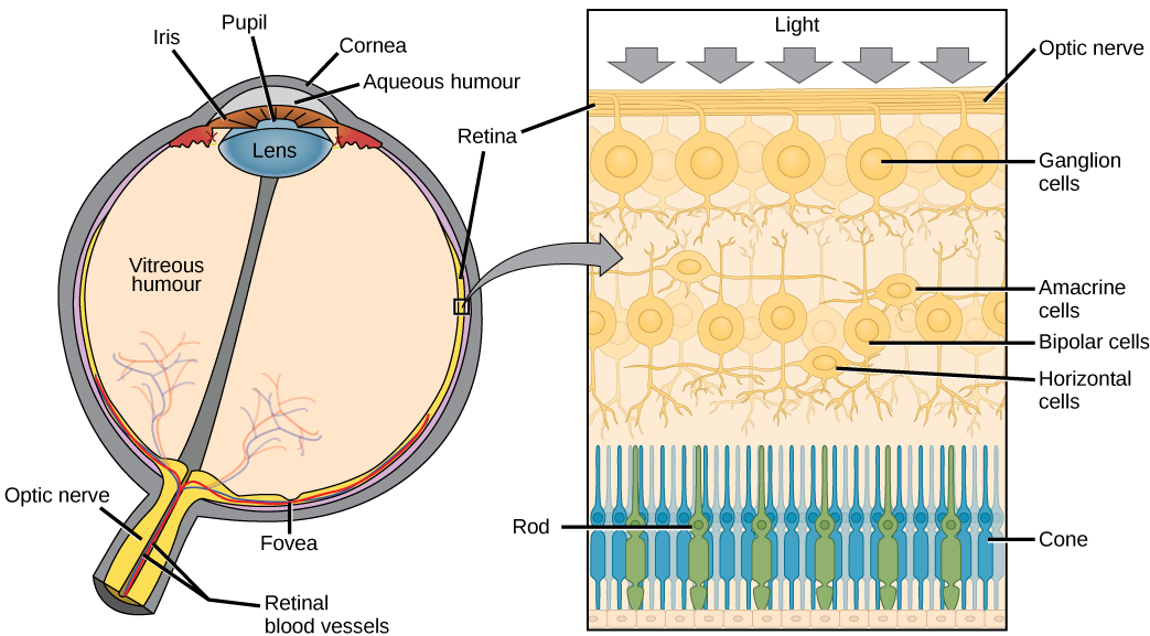
The photoreceptive cells of the eye, where transduction of light to nervous impulses occurs, are located in the retina (shown in [link] ) on the inner surface of the back of the eye. But light does not impinge on the retina unaltered. It passes through other layers that process it so that it can be interpreted by the retina ( [link] b ). The cornea , the front transparent layer of the eye, and the crystalline lens , a transparent convex structure behind the cornea, both refract (bend) light to focus the image on the retina. The iris , which is conspicuous as the colored part of the eye, is a circular muscular ring lying between the lens and cornea that regulates the amount of light entering the eye. In conditions of high ambient light, the iris contracts, reducing the size of the pupil at its center. In conditions of low light, the iris relaxes and the pupil enlarges.

The main function of the lens is to focus light on the retina and fovea centralis. The lens is dynamic, focusing and re-focusing light as the eye rests on near and far objects in the visual field. The lens is operated by muscles that stretch it flat or allow it to thicken, changing the focal length of light coming through it to focus it sharply on the retina. With age comes the loss of the flexibility of the lens, and a form of farsightedness called presbyopia results. Presbyopia occurs because the image focuses behind the retina. Presbyopia is a deficit similar to a different type of farsightedness called hyperopia caused by an eyeball that is too short. For both defects, images in the distance are clear but images nearby are blurry. Myopia (nearsightedness) occurs when an eyeball is elongated and the image focus falls in front of the retina. In this case, images in the distance are blurry but images nearby are clear.
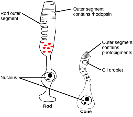
There are two types of photoreceptors in the retina: rods and cones , named for their general appearance as illustrated in [link] . Rods are strongly photosensitive and are located in the outer edges of the retina. They detect dim light and are used primarily for peripheral and nighttime vision. Cones are weakly photosensitive and are located near the center of the retina. They respond to bright light, and their primary role is in daytime, color vision.

The fovea is the region in the center back of the eye that is responsible for acute vision. The fovea has a high density of just cones. When you bring your gaze to an object to examine it intently in bright light, the eyes orient so that the object’s image falls on the fovea. This is the area of the retina that gives us high clarity of vision. However, when looking at a star in the night sky or other object in dim light, the object can be better viewed by the peripheral vision because it is the rods in higher concentrations in the other regions of the retina, rather than the cones at the center, that operate better in low light. In low-light conditions, the rods allow us to see in shades of gray because cones require bright light to be stimulated and don't respond in low light conditions.

Notification Switch
Would you like to follow the 'Human biology' conversation and receive update notifications?