<< Chapter < Page Chapter >> Page >
By the end of this section, you will be able to:
  • Identify the anatomical features of a bone
  • Describe the histology of bone tissue
  • Compare and contrast compact and spongy bone
  • Identify the structures that compose compact and spongy bone
  • Describe how bones are nourished and innervated

Bone tissue (osseous tissue) differs greatly from other tissues in the body. Bone is hard and many of its functions depend on that characteristic hardness. Later discussions in this chapter will show that bone is also dynamic in that its shape adjusts to accommodate stresses. This section will examine the gross anatomy of bone first and then move on to its histology.

Gross anatomy of bone

The structure of a long bone allows for the best visualization of all of the parts of a bone ( [link] ). A long bone has two parts: the diaphysis    and the epiphysis    . The diaphysis is the tubular shaft that runs between the proximal and distal ends of the bone. The hollow region in the diaphysis is called the medullary cavity    , which is filled with yellow marrow. The walls of the diaphysis are composed of dense and hard compact bone    .
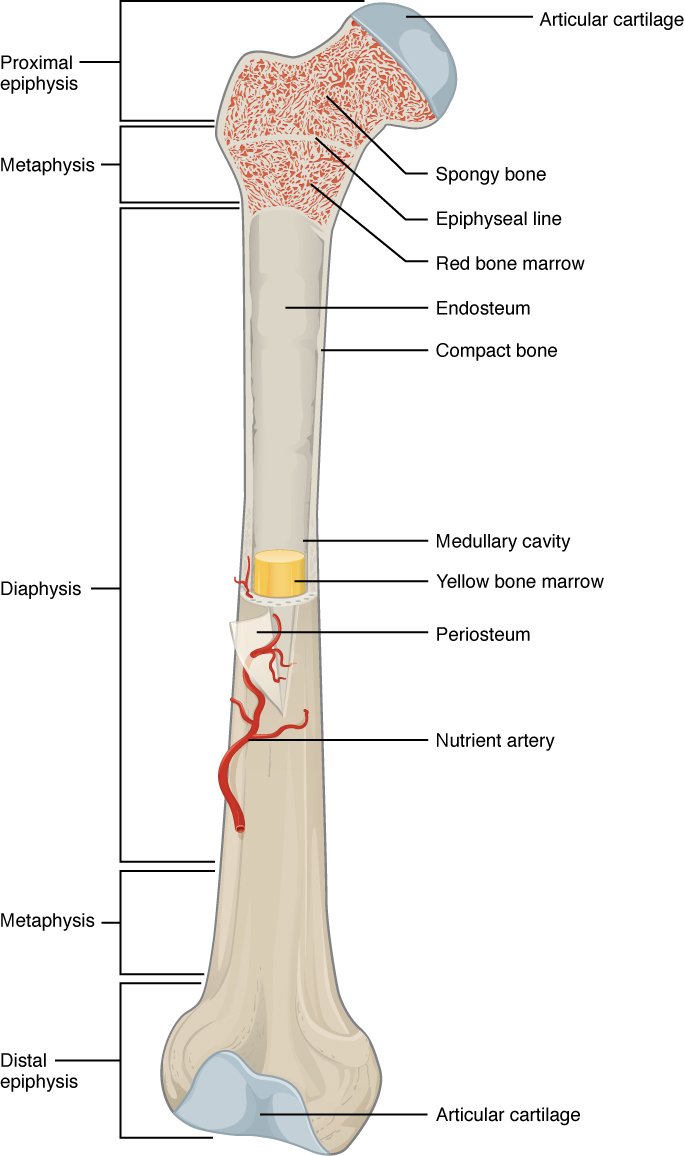
Anatomy of a long bone

This illustration depicts an anterior view of the right femur, or thigh bone. The inferior end that connects to the knee is at the bottom of the diagram and the superior end that connects to the hip is at the top of the diagram. The bottom end of the bone contains a smaller lateral bulge and a larger medial bulge. A blue articular cartilage covers the inner half of each bulge as well as the small trench that runs between the bulges. This area of the inferior end of the bone is labeled the distal epiphysis. Above the distal epiphysis is the metaphysis, where the bone tapers from the wide epiphysis into the relatively thin shaft. The entire length of the shaft is the diaphysis. The superior half of the femur is cut away to show its internal contents. The bone is covered with an outer translucent sheet called the periosteum. At the midpoint of the diaphysis, a nutrient artery travels through the periosteum and into the inner layers of the bone. The periosteum surrounds a white cylinder of solid bone labeled compact bone. The cavity at the center of the compact bone is called the medullary cavity. The inner layer of the compact bone that lines the medullary cavity is called the endosteum. Within the diaphysis, the medullary cavity contains a cylinder of yellow bone marrow that is penetrated by the nutrient artery. The superior end of the femur is also connected to the diaphysis by a metaphysis. In this upper metaphysis, the bone gradually widens between the diaphysis and the proximal epiphysis. The proximal epiphysis of the femur is roughly hexagonal in shape. However, the upper right side of the hexagon has a large, protruding knob. The femur connects and rotates within the hip socket at this knob. The knob is covered with a blue colored articular cartilage. The internal anatomy of the upper metaphysis and proximal epiphysis are revealed. The medullary cavity in these regions is filled with the mesh like spongy bone. Red bone marrow occupies the many cavities within the spongy bone. There is a clear, white line separating the spongy bone of the upper metaphysis with that of the proximal epiphysis. This line is labeled the epiphyseal line.
A typical long bone shows the gross anatomical characteristics of bone.

The wider section at each end of the bone is called the epiphysis (plural = epiphyses), which is filled with spongy bone. Red marrow fills the spaces in the spongy bone. Each epiphysis meets the diaphysis at the metaphysis, the narrow area that contains the epiphyseal plate    (growth plate), a layer of hyaline (transparent) cartilage in a growing bone. When the bone stops growing in early adulthood (approximately 18–21 years), the cartilage is replaced by osseous tissue and the epiphyseal plate becomes an epiphyseal line.

Bone cells and tissue

Bone contains a relatively small number of cells entrenched in a matrix of collagen fibers that provide a surface for inorganic salt crystals to adhere. These salt crystals form when calcium phosphate and calcium carbonate combine to create hydroxyapatite, which incorporates other inorganic salts like magnesium hydroxide, fluoride, and sulfate as it crystallizes, or calcifies, on the collagen fibers. The hydroxyapatite crystals give bones their hardness and strength, while the collagen fibers give them flexibility so that they are not brittle.

Although bone cells compose a small amount of the bone volume, they are crucial to the function of bones. Four types of cells are found within bone tissue: osteoblasts, osteocytes, osteogenic cells, and osteoclasts ( [link] ).
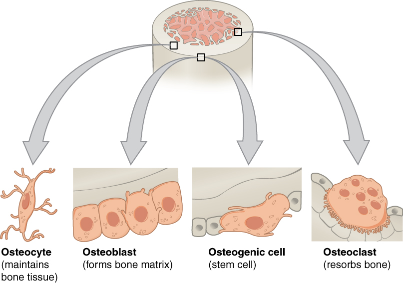
Bone cells

The top of this diagram shows the cross section of a generic bone with three zoom in boxes. The first box is on the periosteum. The second box is on the middle of the compact bone layer. The third box is on the inner edge of the compact bone where it transitions into the spongy bone. The callout in the periosteum points to two images. In the first image, four osteoblast cells are sitting end to end on the periosteum. The osteoblasts are roughly square shaped, except for one of the cells which is developing small, finger like projections. The caption says, “Osteoblasts form the matrix of the bone.” The second image called out from the periosteum shows a large, amorphous osteogenic cell sitting on the periosteum. The osteogenic cell is surrounded on both sides by a row of much smaller osteoblasts. The cell is shaped like a mushroom cap and also has finger like projections. The cell is a stem cell that develops into other bone cells. The box in the middle of the compact bone layer is pointing to an osteocyte. The osteocyte is a thin cell, roughly diamond shaped, with many branching, finger-like projections. The osteoctyes maintain bone tissue. The box at the inner edge of the compact bone is pointing to an osteoclast. The osteoclast is a large, round cell with multiple nuclei. It also has rows of fine finger like projections on its lower surface where it is sitting on the compact bone. The osteoclast reabsorbs bone.
Four types of cells are found within bone tissue. Osteogenic cells are undifferentiated and develop into osteoblasts. When osteoblasts get trapped within the calcified matrix, their structure and function changes, and they become osteocytes. Osteoclasts develop from monocytes and macrophages and differ in appearance from other bone cells.

The osteoblast    is the bone cell responsible for forming new bone and is found in the growing portions of bone, including the periosteum and endosteum. Osteoblasts, which do not divide, synthesize and secrete the collagen matrix and calcium salts. As the secreted matrix surrounding the osteoblast calcifies, the osteoblast become trapped within it; as a result, it changes in structure and becomes an osteocyte    , the primary cell of mature bone and the most common type of bone cell. Each osteocyte is located in a space called a lacuna and is surrounded by bone tissue. Osteocytes maintain the mineral concentration of the matrix via the secretion of enzymes. Like osteoblasts, osteocytes lack mitotic activity. They can communicate with each other and receive nutrients via long cytoplasmic processes that extend through canaliculi    (singular = canaliculus), channels within the bone matrix.

Questions & Answers

what is microbiology
Agebe Reply
What is a cell
Odelana Reply
what is cell
Mohammed
how does Neisseria cause meningitis
Nyibol Reply
what is microbiologist
Muhammad Reply
what is errata
Muhammad
is the branch of biology that deals with the study of microorganisms.
Ntefuni Reply
What is microbiology
Mercy Reply
studies of microbes
Louisiaste
when we takee the specimen which lumbar,spin,
Ziyad Reply
How bacteria create energy to survive?
Muhamad Reply
Bacteria doesn't produce energy they are dependent upon their substrate in case of lack of nutrients they are able to make spores which helps them to sustain in harsh environments
_Adnan
But not all bacteria make spores, l mean Eukaryotic cells have Mitochondria which acts as powerhouse for them, since bacteria don't have it, what is the substitution for it?
Muhamad
they make spores
Louisiaste
what is sporadic nd endemic, epidemic
Aminu Reply
the significance of food webs for disease transmission
Abreham
food webs brings about an infection as an individual depends on number of diseased foods or carriers dully.
Mark
explain assimilatory nitrate reduction
Esinniobiwa Reply
Assimilatory nitrate reduction is a process that occurs in some microorganisms, such as bacteria and archaea, in which nitrate (NO3-) is reduced to nitrite (NO2-), and then further reduced to ammonia (NH3).
Elkana
This process is called assimilatory nitrate reduction because the nitrogen that is produced is incorporated in the cells of microorganisms where it can be used in the synthesis of amino acids and other nitrogen products
Elkana
Examples of thermophilic organisms
Shu Reply
Give Examples of thermophilic organisms
Shu
advantages of normal Flora to the host
Micheal Reply
Prevent foreign microbes to the host
Abubakar
they provide healthier benefits to their hosts
ayesha
They are friends to host only when Host immune system is strong and become enemies when the host immune system is weakened . very bad relationship!
Mark
what is cell
faisal Reply
cell is the smallest unit of life
Fauziya
cell is the smallest unit of life
Akanni
ok
Innocent
cell is the structural and functional unit of life
Hasan
is the fundamental units of Life
Musa
what are emergency diseases
Micheal Reply
There are nothing like emergency disease but there are some common medical emergency which can occur simultaneously like Bleeding,heart attack,Breathing difficulties,severe pain heart stock.Hope you will get my point .Have a nice day ❣️
_Adnan
define infection ,prevention and control
Innocent
I think infection prevention and control is the avoidance of all things we do that gives out break of infections and promotion of health practices that promote life
Lubega
Heyy Lubega hussein where are u from?
_Adnan
en français
Adama
which site have a normal flora
ESTHER Reply
Many sites of the body have it Skin Nasal cavity Oral cavity Gastro intestinal tract
Safaa
skin
Asiina
skin,Oral,Nasal,GIt
Sadik
How can Commensal can Bacteria change into pathogen?
Sadik
How can Commensal Bacteria change into pathogen?
Sadik
all
Tesfaye
by fussion
Asiina
what are the advantages of normal Flora to the host
Micheal
what are the ways of control and prevention of nosocomial infection in the hospital
Micheal
Got questions? Join the online conversation and get instant answers!
Jobilize.com Reply

Get Jobilize Job Search Mobile App in your pocket Now!

Get it on Google Play Download on the App Store Now


Source:  OpenStax, Human biology. OpenStax CNX. Dec 01, 2015 Download for free at http://legacy.cnx.org/content/col11903/1.3
Google Play and the Google Play logo are trademarks of Google Inc.

Notification Switch

Would you like to follow the 'Human biology' conversation and receive update notifications?

Ask