<< Chapter < Page Chapter >> Page >
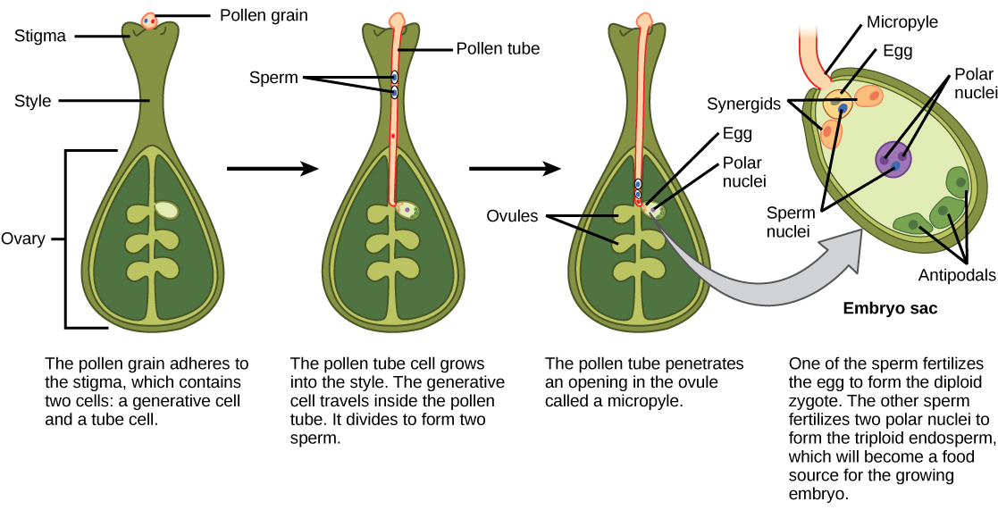
 Illustration shows the gynoecium of a flowering plant. A pollen grain adheres to the stigma. The pollen contains two cells: a generative cell and a tube cell. The pollen tube cell grows into the style. The generative cell travels inside the pollen tube. It divides to form two sperm. The pollen tube penetrates an opening in the ovule called a micropyle. One of the sperm fertilizes the egg to form the zygote. The other sperm fertilizes two polar nuclei to form a triploid endosperm, which becomes a food source for the growing embryo.
In angiosperms, one sperm fertilizes the egg to form the 2 n zygote, and the other sperm fertilizes the central cell to form the 3 n endosperm. This is called a double fertilization.

After fertilization, the zygote divides to form two cells: the upper cell, or terminal cell, and the lower, or basal, cell. The division of the basal cell gives rise to the suspensor    , which eventually makes connection with the maternal tissue. The suspensor provides a route for nutrition to be transported from the mother plant to the growing embryo. The terminal cell also divides, giving rise to a globular-shaped proembryo ( [link] a ). In dicots (eudicots), the developing embryo has a heart shape, due to the presence of the two rudimentary cotyledons ( [link] b ). In non-endospermic dicots, such as Capsella bursa , the endosperm develops initially, but is then digested, and the food reserves are moved into the two cotyledons. As the embryo and cotyledons enlarge, they run out of room inside the developing seed, and are forced to bend ( [link] c ). Ultimately, the embryo and cotyledons fill the seed ( [link] d ), and the seed is ready for dispersal. Embryonic development is suspended after some time, and growth is resumed only when the seed germinates. The developing seedling will rely on the food reserves stored in the cotyledons until the first set of leaves begin photosynthesis.
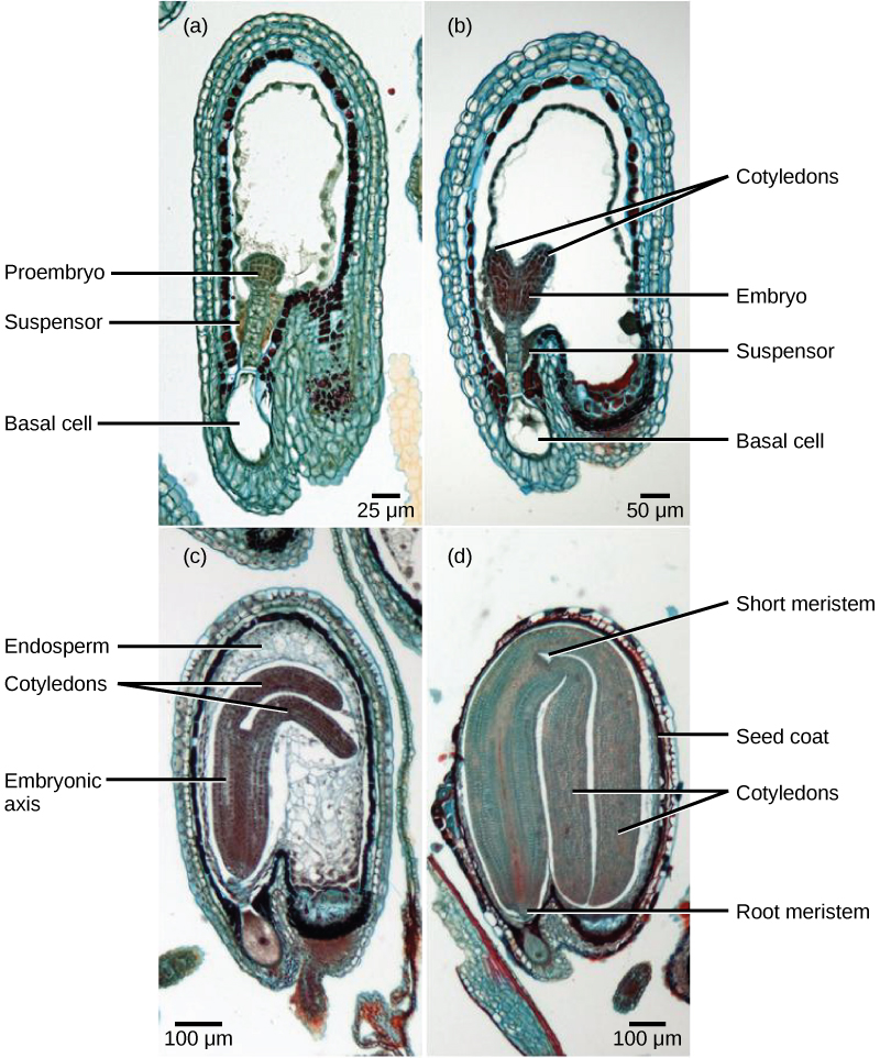
 Micrograph A shows a seed in the initial stage of development. The proembryo grows inside an oval-shaped ovary with an opening at the bottom. The basal cell is at the bottom ovary, and suspensor cells are above it. The globular proembryo grows at the top of the suspensor. Micrograph B shows the second stage of development, in which the embryo grows into a heart-shape. Each bump in the heart is a cotyledon. Micrograph C shows the third stage of development. The embryo has grown longer and wider, and the cotyledons have grown into long extensions resembling bunny ears bent so they fit inside the seed. Cells inside the embryo grow in vertical columns. The central column, between the two ears, is called the embryonic axis. Micrograph D shows the fourth stage of development. The bunny ears are now as large as the main part of the embryo, and completely folded over. The base of the embryo is the root meristem, and the space between the two ears is the shoot meristem. A seed coat has formed over the ovary.
Shown are the stages of embryo development in the ovule of a shepherd’s purse ( Capsella bursa ). After fertilization, the zygote divides to form an upper terminal cell and a lower basal cell. (a) In the first stage of development, the terminal cell divides, forming a globular pro-embryo. The basal cell also divides, giving rise to the suspensor. (b) In the second stage, the developing embryo has a heart shape due to the presence of cotyledons. (c) In the third stage, the growing embryo runs out of room and starts to bend. (d) Eventually, it completely fills the seed. (credit: modification of work by Robert R. Wise; scale-bar data from Matt Russell)

Development of the seed

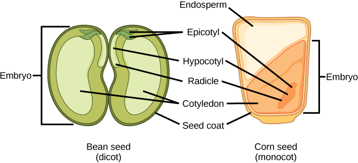
The mature ovule develops into the seed. A typical seed contains a seed coat, cotyledons, endosperm, and a single embryo ( [link] ).
Art connection

 Illustration shows the structure of a monocot corn seed and a dicot bean seed. The lower half of the monocot seed contains the cotyledon, and the upper half contains the endosperm. The dicot seed does not contain an endosperm, but has two cotyledons, one on each side of the bean. Both the monocot and the dicot seed have an epicotyl that is attached to a hypocotyl. The hypocotyl terminates in a radicle. In the dicot, the epicotyl is in the upper middle part of the seed. In the monocot, the epicotyl is in the lower cotyledon. Both the monocot and dicot seed are surrounded by a seed coat.
The structures of dicot and monocot seeds are shown. Dicots (left) have two cotyledons. Monocots, such as corn (right), have one cotyledon, called the scutellum; it channels nutrition to the growing embryo. Both monocot and dicot embryos have a plumule that forms the leaves, a hypocotyl that forms the stem, and a radicle that forms the root. The embryonic axis comprises everything between the plumule and the radicle, not including the cotyledon(s).

What is of the following statements is true?

  1. Both monocots and dicots have an endosperm.
  2. The radicle develops into the root.
  3. The plumule is part of the epicotyl
  4. The endosperm is part of the embryo.

The storage of food reserves in angiosperm seeds differs between monocots and dicots. In monocots, such as corn and wheat, the single cotyledon is called a scutellum    ; the scutellum is connected directly to the embryo via vascular tissue (xylem and phloem). Food reserves are stored in the large endosperm. Upon germination, enzymes are secreted by the aleurone    , a single layer of cells just inside the seed coat that surrounds the endosperm and embryo. The enzymes degrade the stored carbohydrates, proteins and lipids, the products of which are absorbed by the scutellum and transported via a vasculature strand to the developing embryo. Therefore, the scutellum can be seen to be an absorptive organ, not a storage organ.

Questions & Answers

what are components of cells
ofosola Reply
twugzfisfjxxkvdsifgfuy7 it
Sami
58214993
Sami
what is a salt
John
the difference between male and female reproduction
John
what is computed
IBRAHIM Reply
what is biology
IBRAHIM
what is the full meaning of biology
IBRAHIM
what is biology
Jeneba
what is cell
Kuot
425844168
Sami
what is cytoplasm
Emmanuel Reply
structure of an animal cell
Arrey Reply
what happens when the eustachian tube is blocked
Puseletso Reply
what's atoms
Achol Reply
discuss how the following factors such as predation risk, competition and habitat structure influence animal's foraging behavior in essay form
Burnet Reply
cell?
Kuot
location of cervical vertebra
KENNEDY Reply
What are acid
Sheriff Reply
define biology infour way
Happiness Reply
What are types of cell
Nansoh Reply
how can I get this book
Gatyin Reply
what is lump
Chineye Reply
what is cell
Maluak Reply
what is biology
Maluak
what is vertibrate
Jeneba
what's cornea?
Majak Reply
what are cell
Achol

Get Jobilize Job Search Mobile App in your pocket Now!

Get it on Google Play Download on the App Store Now


Source:  OpenStax, Biology. OpenStax CNX. Feb 29, 2016 Download for free at http://cnx.org/content/col11448/1.10
Google Play and the Google Play logo are trademarks of Google Inc.

Notification Switch

Would you like to follow the 'Biology' conversation and receive update notifications?

Ask