<< Chapter < Page Chapter >> Page >

Art connection

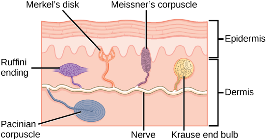
Illustration shows the location of various mechanoreceptors in a cross section of the epidermis and dermis. A nerve runs along the middle of the dermis, and all the mechanoreceptors are connected to it. Ruffini endings, Merkel’s disks, and Meissner’s corpuscles are all located in the upper dermis above the nerve. Ruffini endings are bulbous, horizontal mechanoreceptors located in the middle of the upper dermis. Meissner’s corpuscles are bulbous, vertical mechanoreceptors that touch the bottom of the epidermis. Merkel’s disks have finger-like projections that also touch the bottom of the epidermis. The last type of mechanoreceptor, Pacini corpuscles, are oval mechanoreceptors located in the lower dermis.
Four of the primary mechanoreceptors in human skin are shown. Merkel’s disks, which are unencapsulated, respond to light touch. Meissner’s corpuscles, Ruffini endings, Pacinian corpuscles, and Krause end bulbs are all encapsulated. Meissner’s corpuscles respond to touch and low-frequency vibration. Ruffini endings detect stretch, deformation within joints, and warmth. Pacinian corpuscles detect transient pressure and high-frequency vibration. Krause end bulbs detect cold.

Which of the following statements about mechanoreceptors is false?

  1. Pacini corpuscles are found in both glabrous and hairy skin.
  2. Merkel’s disks are abundant on the fingertips and lips.
  3. Ruffini endings are encapsulated mechanoreceptors.
  4. Meissner’s corpuscles extend into the lower dermis.

Meissner’s corpuscles , (shown in [link] ) also known as tactile corpuscles, are found in the upper dermis, but they project into the epidermis. They, too, are found primarily in the glabrous skin on the fingertips and eyelids. They respond to fine touch and pressure, but they also respond to low-frequency vibration or flutter. They are rapidly adapting, fluid-filled, encapsulated neurons with small, well-defined borders and are responsive to fine details. Like Merkel’s disks, Meissner’s corpuscles are not as plentiful in the palms as they are in the fingertips.
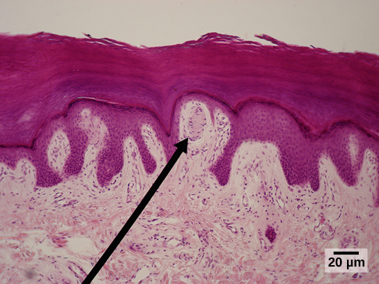
Micrograph shows the epidermis, which stains dark pink and the dermis, which stains light pink. Finger-like projections of epidermis extend into the dermis. Between two of these fingers is an oval Meissner corpuscle about ten microns across and 20 microns long.
Meissner corpuscles in the fingertips, such as the one viewed here using bright field light microscopy, allow for touch discrimination of fine detail. (credit: modification of work by "Wbensmith"/Wikimedia Commons; scale-bar data from Matt Russell)

Deeper in the epidermis, near the base, are Ruffini endings , which are also known as bulbous corpuscles. They are found in both glabrous and hairy skin. These are slow-adapting, encapsulated mechanoreceptors that detect skin stretch and deformations within joints, so they provide valuable feedback for gripping objects and controlling finger position and movement. Thus, they also contribute to proprioception and kinesthesia. Ruffini endings also detect warmth. Note that these warmth detectors are situated deeper in the skin than are the cold detectors. It is not surprising, then, that humans detect cold stimuli before they detect warm stimuli.

Pacinian corpuscles (seen in [link] ) are located deep in the dermis of both glabrous and hairy skin and are structurally similar to Meissner’s corpuscles; they are found in the bone periosteum, joint capsules, pancreas and other viscera, breast, and genitals. They are rapidly adapting mechanoreceptors that sense deep transient (but not prolonged) pressure and high-frequency vibration. Pacinian receptors detect pressure and vibration by being compressed, stimulating their internal dendrites. There are fewer Pacinian corpuscles and Ruffini endings in skin than there are Merkel’s disks and Meissner’s corpuscles.
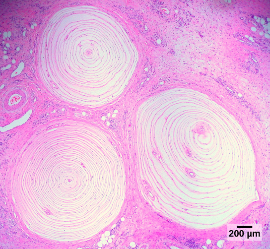
Micrograph shows three Pacinian corpuscles embedded in the dermis. The corpuscles are round and about 1.4 millimeters across and have rings, like a tree stump.
Pacinian corpuscles, such as these visualized using bright field light microscopy, detect pressure (touch) and high-frequency vibration. (credit: modification of work by Ed Uthman; scale-bar data from Matt Russell)

Questions & Answers

what are components of cells
ofosola Reply
twugzfisfjxxkvdsifgfuy7 it
Sami
58214993
Sami
what is a salt
John
the difference between male and female reproduction
John
what is computed
IBRAHIM Reply
what is biology
IBRAHIM
what is the full meaning of biology
IBRAHIM
what is biology
Jeneba
what is cell
Kuot
425844168
Sami
what is cytoplasm
Emmanuel Reply
structure of an animal cell
Arrey Reply
what happens when the eustachian tube is blocked
Puseletso Reply
what's atoms
Achol Reply
discuss how the following factors such as predation risk, competition and habitat structure influence animal's foraging behavior in essay form
Burnet Reply
cell?
Kuot
location of cervical vertebra
KENNEDY Reply
What are acid
Sheriff Reply
define biology infour way
Happiness Reply
What are types of cell
Nansoh Reply
how can I get this book
Gatyin Reply
what is lump
Chineye Reply
what is cell
Maluak Reply
what is biology
Maluak
what is vertibrate
Jeneba
what's cornea?
Majak Reply
what are cell
Achol

Get Jobilize Job Search Mobile App in your pocket Now!

Get it on Google Play Download on the App Store Now


Source:  OpenStax, Biology. OpenStax CNX. Feb 29, 2016 Download for free at http://cnx.org/content/col11448/1.10
Google Play and the Google Play logo are trademarks of Google Inc.

Notification Switch

Would you like to follow the 'Biology' conversation and receive update notifications?

Ask