<< Chapter < Page Chapter >> Page >

Leakage channels

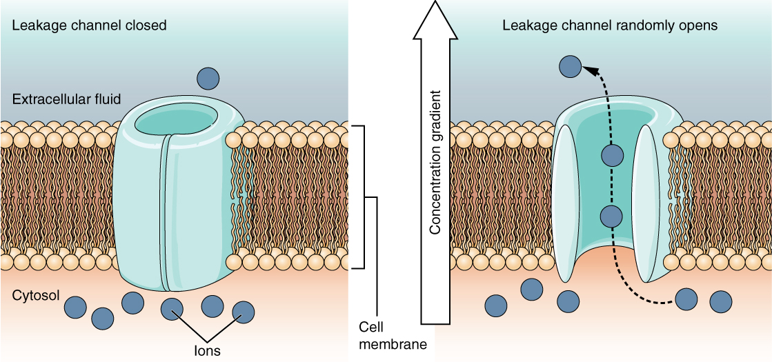
This is a two part diagram. Both diagrams show a leakage channel embedded in the lipid membrane bilayer. The leakage channel is cylindrical with a large, central opening. In the first diagram there are several ions in the cytosol but only one ion in the extracellular fluid. No ions are moving through the leakage channel because the channel is closed. In the second diagram, the leakage channel randomly opens, allowing two ions to travel through the channel, down their concentration gradient, and out into the extracellular fluid.
In certain situations, ions need to move across the membrane randomly. The particular electrical properties of certain cells are modified by the presence of this type of channel.

The membrane potential

The electrical state of the cell membrane can have several variations. These are all variations in the membrane potential    . A potential is a distribution of charge across the cell membrane, measured in millivolts (mV). The standard is to compare the inside of the cell relative to the outside, so the membrane potential is a value representing the charge on the intracellular side of the membrane based on the outside being zero, relatively speaking ( [link] ).
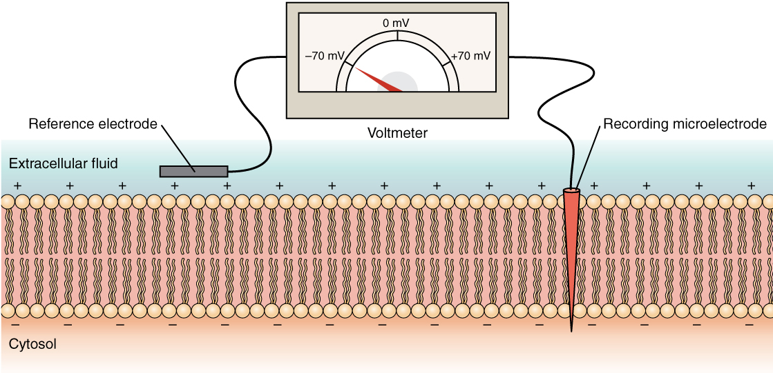
Measuring charge across a membrane with a voltmeter

This diagram shows a cross section of a cell membrane. The extracellular fluid side of the cell membrane is positively charged while the cytosol side of the membrane is negatively charged. There is a microelectrode embedded in the cell membrane. The microelectrode is attached to a voltmeter, which also has a reference electrode on the extracellular fluid side. The readout of the voltmeter is negative 70 millivolts.
A recording electrode is inserted into the cell and a reference electrode is outside the cell. By comparing the charge measured by these two electrodes, the transmembrane voltage is determined. It is conventional to express that value for the cytosol relative to the outside.

The concentration of ions in extracellular and intracellular fluids is largely balanced, with a net neutral charge. However, a slight difference in charge occurs right at the membrane surface, both internally and externally. It is the difference in this very limited region that has all the power in neurons (and muscle cells) to generate electrical signals, including action potentials.

Before these electrical signals can be described, the resting state of the membrane must be explained. When the cell is at rest, and the ion channels are closed (except for leakage channels which randomly open), ions are distributed across the membrane in a very predictable way. The concentration of Na + outside the cell is 10 times greater than the concentration inside. Also, the concentration of K + inside the cell is greater than outside. The cytosol contains a high concentration of anions, in the form of phosphate ions and negatively charged proteins. Large anions are a component of the inner cell membrane, including specialized phospholipids and proteins associated with the inner leaflet of the membrane (leaflet is a term used for one side of the lipid bilayer membrane). The negative charge is localized in the large anions.

With the ions distributed across the membrane at these concentrations, the difference in charge is measured at -70 mV, the value described as the resting membrane potential    . The exact value measured for the resting membrane potential varies between cells, but -70 mV is most commonly used as this value. This voltage would actually be much lower except for the contributions of some important proteins in the membrane. Leakage channels allow Na + to slowly move into the cell or K + to slowly move out, and the Na + /K + pump restores them. This may appear to be a waste of energy, but each has a role in maintaining the membrane potential.

The action potential

Resting membrane potential describes the steady state of the cell, which is a dynamic process that is balanced by ion leakage and ion pumping. Without any outside influence, it will not change. To get an electrical signal started, the membrane potential has to change.

Questions & Answers

what is hypogelersomia
aliyu Reply
what are the parts of the female reproductive system?
Orji Reply
what is anatomy
Divinefavour Reply
what are the six types of synovial joints and their ligaments
Darlington Reply
draw the six types of synovial joint and their ligaments
Darlington
System of human beings
Katumi Reply
System in humans body
Katumi
Diagram of animals and plants cell
Favour Reply
at what age does development of bone end
Alal Reply
how many bones are in the human upper layers
Daniel Reply
how many bones do we have
Nbeke
bones that form the wrist
Priscilla Reply
yes because it is in the range of neutrophil count
Alexander Reply
because their basic work is to fight against harmful external bodies and they are always present when chematoxin are released in an area in body
Alexander
What is pathology
Samuel Reply
what is pathology
Nbeke
what's pathology
Nbeke
what is anatomy
ESTHER Reply
drowning and level female reproductive system
Anas Reply
what are the types of homeostasis
Odey Reply
diagram of the digestive system
Zainab Reply
drown and level female reproductive system
Anas
anatomy
Anas

Get Jobilize Job Search Mobile App in your pocket Now!

Get it on Google Play Download on the App Store Now


Source:  OpenStax, Anatomy & Physiology. OpenStax CNX. Feb 04, 2016 Download for free at http://legacy.cnx.org/content/col11496/1.8
Google Play and the Google Play logo are trademarks of Google Inc.

Notification Switch

Would you like to follow the 'Anatomy & Physiology' conversation and receive update notifications?

Ask