| << Chapter < Page | Chapter >> Page > |

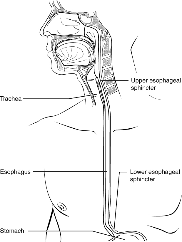
The mucosa of the esophagus is made up of an epithelial lining that contains non-keratinized, stratified squamous epithelium, with a layer of basal and parabasal cells. This epithelium protects against erosion from food particles. The mucosa’s lamina propria contains mucus-secreting glands. The muscularis layer changes according to location: In the upper third of the esophagus, the muscularis is skeletal muscle. In the middle third, it is both skeletal and smooth muscle. In the lower third, it is smooth muscle. As mentioned previously, the most superficial layer of the esophagus is called the adventitia, not the serosa. In contrast to the stomach and intestines, the loose connective tissue of the adventitia is not covered by a fold of visceral peritoneum. The digestive functions of the esophagus are identified in [link] .
| Digestive Functions of the Esophagus | |
|---|---|
| Action | Outcome |
| Upper esophageal sphincter relaxation | Allows the bolus to move from the laryngopharynx to the esophagus |
| Peristalsis | Propels the bolus through the esophagus |
| Lower esophageal sphincter relaxation | Allows the bolus to move from the esophagus into the stomach and prevents chime from entering the esophagus |
| Mucus secretion | Lubricates the esophagus, allowing easy passage of the bolus |
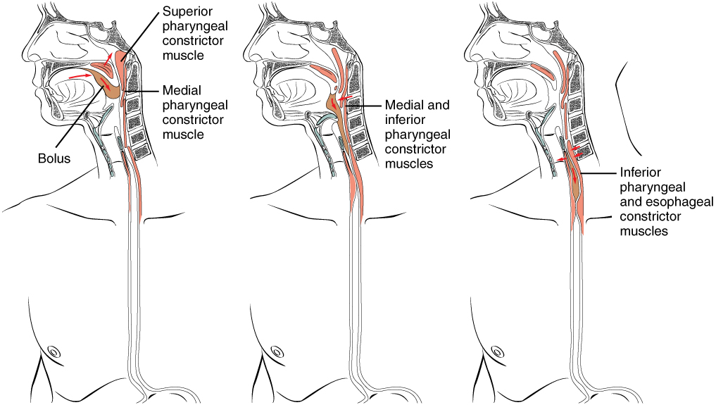
Deglutition is another word for swallowing—the movement of food from the mouth to the stomach. The entire process takes about 4 to 8 seconds for solid or semisolid food, and about 1 second for very soft food and liquids. Although this sounds quick and effortless, deglutition is, in fact, a complex process that involves both the skeletal muscle of the tongue and the muscles of the pharynx and esophagus. It is aided by the presence of mucus and saliva. There are three stages in deglutition: the voluntary phase, the pharyngeal phase, and the esophageal phase ( [link] ). The autonomic nervous system controls the latter two phases.

The voluntary phase of deglutition (also known as the oral or buccal phase) is so called because you can control when you swallow food. In this phase, chewing has been completed and swallowing is set in motion. The tongue moves upward and backward against the palate, pushing the bolus to the back of the oral cavity and into the oropharynx. Other muscles keep the mouth closed and prevent food from falling out. At this point, the two involuntary phases of swallowing begin.
In the pharyngeal phase, stimulation of receptors in the oropharynx sends impulses to the deglutition center (a collection of neurons that controls swallowing) in the medulla oblongata. Impulses are then sent back to the uvula and soft palate, causing them to move upward and close off the nasopharynx. The laryngeal muscles also constrict to prevent aspiration of food into the trachea. At this point, deglutition apnea takes place, which means that breathing ceases for a very brief time. Contractions of the pharyngeal constrictor muscles move the bolus through the oropharynx and laryngopharynx. Relaxation of the upper esophageal sphincter then allows food to enter the esophagus.
The entry of food into the esophagus marks the beginning of the esophageal phase of deglutition and the initiation of peristalsis. As in the previous phase, the complex neuromuscular actions are controlled by the medulla oblongata. Peristalsis propels the bolus through the esophagus and toward the stomach. The circular muscle layer of the muscularis contracts, pinching the esophageal wall and forcing the bolus forward. At the same time, the longitudinal muscle layer of the muscularis also contracts, shortening this area and pushing out its walls to receive the bolus. In this way, a series of contractions keeps moving food toward the stomach. When the bolus nears the stomach, distention of the esophagus initiates a short reflex relaxation of the lower esophageal sphincter that allows the bolus to pass into the stomach. During the esophageal phase, esophageal glands secrete mucus that lubricates the bolus and minimizes friction.
Watch this animation to see how swallowing is a complex process that involves the nervous system to coordinate the actions of upper respiratory and digestive activities. During which stage of swallowing is there a risk of food entering respiratory pathways and how is this risk blocked?
In the mouth, the tongue and the teeth begin mechanical digestion, and saliva begins chemical digestion. The pharynx, which plays roles in breathing and vocalization as well as digestion, runs from the nasal and oral cavities superiorly to the esophagus inferiorly (for digestion) and to the larynx anteriorly (for respiration). During deglutition (swallowing), the soft palate rises to close off the nasopharynx, the larynx elevates, and the epiglottis folds over the glottis. The esophagus includes an upper esophageal sphincter made of skeletal muscle, which regulates the movement of food from the pharynx to the esophagus. It also has a lower esophageal sphincter, made of smooth muscle, which controls the passage of food from the esophagus to the stomach. Cells in the esophageal wall secrete mucus that eases the passage of the food bolus.
Watch this animation to see how swallowing is a complex process that involves the nervous system to coordinate the actions of upper respiratory and digestive activities. During which stage of swallowing is there a risk of food entering respiratory pathways and how is this risk blocked?
Answers may vary.
van Loon FPL, Holmes SJ, Sirotkin B, Williams W, Cochi S, Hadler S, Lindegren ML. Morbidity and Mortality Weekly Report: Mumps surveillance -- United States, 1988–1993 [Internet]. Atlanta, GA: Center for Disease Control; [cited 2013 Apr 3]. Available from: (External Link) .

Notification Switch
Would you like to follow the 'Anatomy & Physiology' conversation and receive update notifications?