<< Chapter < Page Chapter >> Page >
By the end of this section, you will be able to:
  • Compare and contrast the three tunics that make up the walls of most blood vessels
  • Distinguish between elastic arteries, muscular arteries, and arterioles on the basis of structure, location, and function
  • Describe the basic structure of a capillary bed, from the supplying metarteriole to the venule into which it drains
  • Explain the structure and function of venous valves in the large veins of the extremities

Blood is carried through the body via blood vessels. An artery is a blood vessel that carries blood away from the heart, where it branches into ever-smaller vessels. Eventually, the smallest arteries, vessels called arterioles, further branch into tiny capillaries, where nutrients and wastes are exchanged, and then combine with other vessels that exit capillaries to form venules, small blood vessels that carry blood to a vein, a larger blood vessel that returns blood to the heart.

Arteries and veins transport blood in two distinct circuits: the systemic circuit and the pulmonary circuit ( [link] ). Systemic arteries provide blood rich in oxygen to the body’s tissues. The blood returned to the heart through systemic veins has less oxygen, since much of the oxygen carried by the arteries has been delivered to the cells. In contrast, in the pulmonary circuit, arteries carry blood low in oxygen exclusively to the lungs for gas exchange. Pulmonary veins then return freshly oxygenated blood from the lungs to the heart to be pumped back out into systemic circulation. Although arteries and veins differ structurally and functionally, they share certain features.
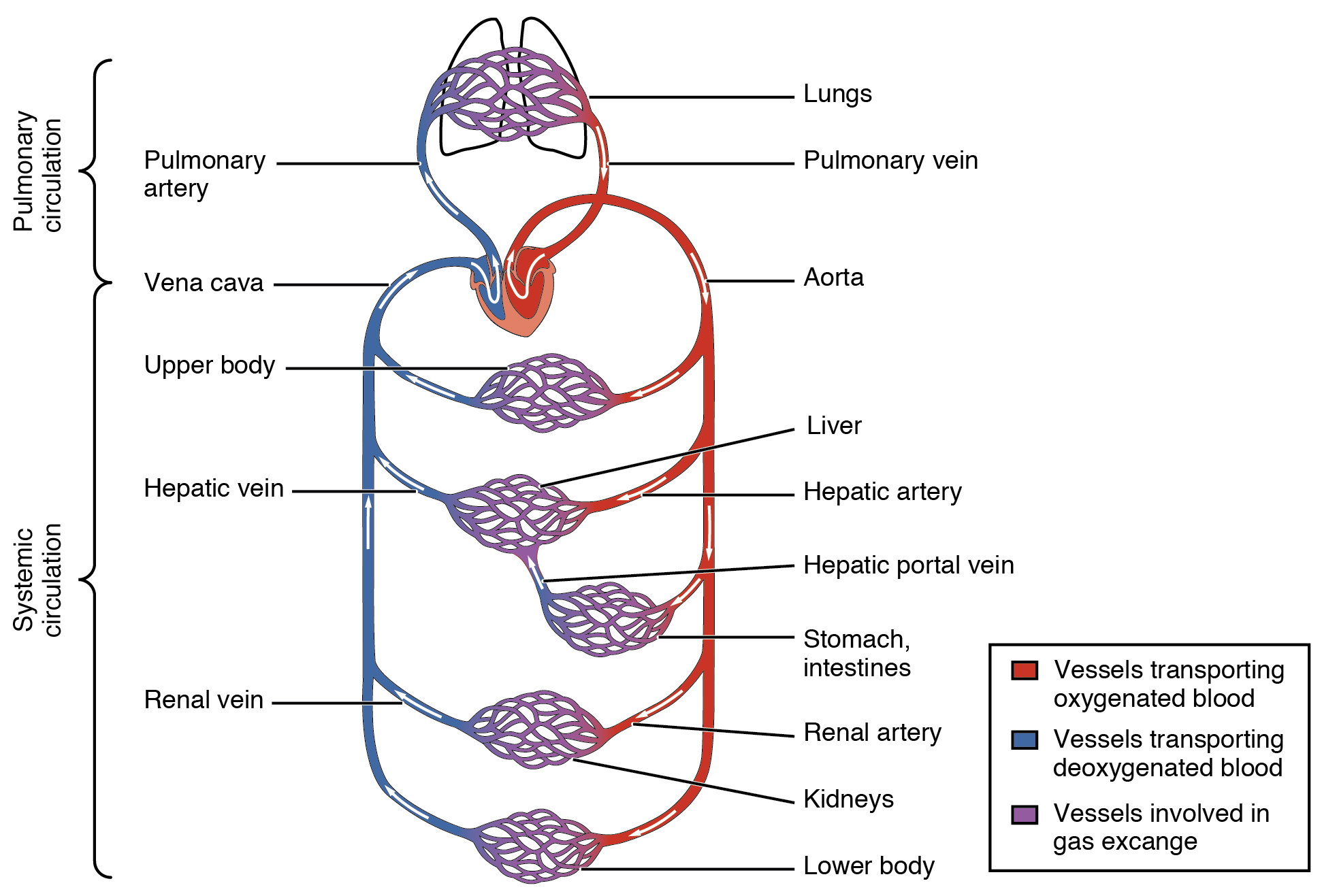
Cardiovascular circulation

This diagram shows how oxygenated and deoxygenated blood flow through the major organs in the body.
The pulmonary circuit moves blood from the right side of the heart to the lungs and back to the heart. The systemic circuit moves blood from the left side of the heart to the head and body and returns it to the right side of the heart to repeat the cycle. The arrows indicate the direction of blood flow, and the colors show the relative levels of oxygen concentration.

Shared structures

Different types of blood vessels vary slightly in their structures, but they share the same general features. Arteries and arterioles have thicker walls than veins and venules because they are closer to the heart and receive blood that is surging at a far greater pressure ( [link] ). Each type of vessel has a lumen    —a hollow passageway through which blood flows. Arteries have smaller lumens than veins, a characteristic that helps to maintain the pressure of blood moving through the system. Together, their thicker walls and smaller diameters give arterial lumens a more rounded appearance in cross section than the lumens of veins.
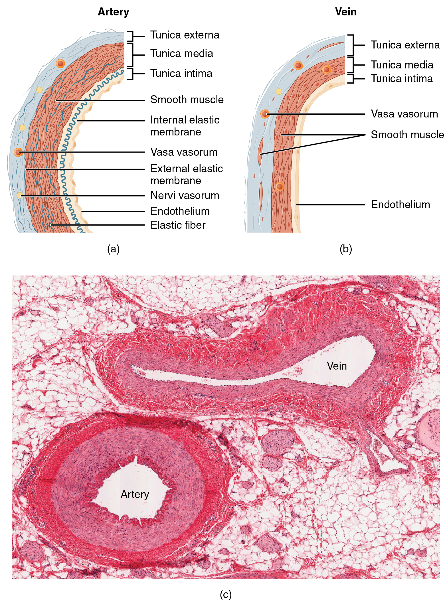
Structure of blood vessels

The top left panel of this figure shows the ultrastructure of an artery, and the top right panel shows the ultrastructure of a vein. The bottom panel shows a micrograph with the cross sections of an artery and a vein.
(a) Arteries and (b) veins share the same general features, but the walls of arteries are much thicker because of the higher pressure of the blood that flows through them. (c) A micrograph shows the relative differences in thickness. LM × 160. (Micrograph provided by the Regents of the University of Michigan Medical School © 2012)

Questions & Answers

what is a blood vessels
Sani Reply
what is plasma and is component
Fad Reply
what is the anterior
Tito Reply
Means front part of the body
Ibrahim
what is anatomy
Ruth Reply
To better understand how the different part of the body works. To understand the physiology of the various structures in the body. To differentiate the systems of the human body .
Roseann Reply
what is hypogelersomia
aliyu Reply
what are the parts of the female reproductive system?
Orji Reply
what is anatomy
Divinefavour Reply
what are the six types of synovial joints and their ligaments
Darlington Reply
draw the six types of synovial joint and their ligaments
Darlington
System of human beings
Katumi Reply
System in humans body
Katumi
Diagram of animals and plants cell
Favour Reply
at what age does development of bone end
Alal Reply
how many bones are in the human upper layers
Daniel Reply
how many bones do we have
Nbeke
bones that form the wrist
Priscilla Reply
yes because it is in the range of neutrophil count
Alexander Reply
because their basic work is to fight against harmful external bodies and they are always present when chematoxin are released in an area in body
Alexander

Get Jobilize Job Search Mobile App in your pocket Now!

Get it on Google Play Download on the App Store Now


Source:  OpenStax, Anatomy & Physiology. OpenStax CNX. Feb 04, 2016 Download for free at http://legacy.cnx.org/content/col11496/1.8
Google Play and the Google Play logo are trademarks of Google Inc.

Notification Switch

Would you like to follow the 'Anatomy & Physiology' conversation and receive update notifications?

Ask