<< Chapter < Page Chapter >> Page >
By the end of this section, you will be able to:
  • Describe the basic structure of a neuron
  • Identify the different types of neurons on the basis of polarity
  • List the glial cells of the CNS and describe their function
  • List the glial cells of the PNS and describe their function

Nervous tissue is composed of two types of cells, neurons and glial cells. Neurons are the primary type of cell that most anyone associates with the nervous system. They are responsible for the computation and communication that the nervous system provides. They are electrically active and release chemical signals to target cells. Glial cells, or glia, are known to play a supporting role for nervous tissue. Ongoing research pursues an expanded role that glial cells might play in signaling, but neurons are still considered the basis of this function. Neurons are important, but without glial support they would not be able to perform their function.

Neurons

Neurons are the cells considered to be the basis of nervous tissue. They are responsible for the electrical signals that communicate information about sensations, and that produce movements in response to those stimuli, along with inducing thought processes within the brain. An important part of the function of neurons is in their structure, or shape. The three-dimensional shape of these cells makes the immense numbers of connections within the nervous system possible.

Parts of a neuron

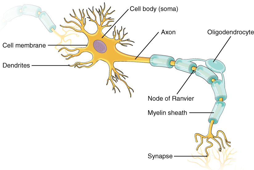
As you learned in the first section, the main part of a neuron is the cell body, which is also known as the soma (soma = “body”). The cell body contains the nucleus and most of the major organelles. But what makes neurons special is that they have many extensions of their cell membranes, which are generally referred to as processes. Neurons are usually described as having one, and only one, axon—a fiber that emerges from the cell body and projects to target cells. That single axon can branch repeatedly to communicate with many target cells. It is the axon that propagates the nerve impulse, which is communicated to one or more cells. The other processes of the neuron are dendrites, which receive information from other neurons at specialized areas of contact called synapses . The dendrites are usually highly branched processes, providing locations for other neurons to communicate with the cell body. Information flows through a neuron from the dendrites, across the cell body, and down the axon. This gives the neuron a polarity—meaning that information flows in this one direction. [link] shows the relationship of these parts to one another.
Parts of a neuron

This illustration shows the anatomy of a neuron. The neuron has a very irregular cell body (soma) containing a purple nucleus. There are six projections protruding from the top, bottom and left side of the cell body. Each of the projections branches many times, forming small, tree-shaped structures protruding from the cell body. The right side of the cell body tapers into a long cord called the axon. The axon is insulated by segments of myelin sheath, which resemble a semitransparent toilet paper roll wound around the axon. The myelin sheath is not continuous, but is separated into equally spaced segments. The bare axon segments between the sheath segments are called nodes of Ranvier. An oligodendrocyte is reaching its two arm like projections onto two myelin sheath segments. The axon branches many times at its end, where it connects to the dendrites of another neuron. Each connection between an axon branch and a dendrite is called a synapse. The cell membrane completely surrounds the cell body, dendrites, and its axon. The axon of another nerve is seen in the upper left of the diagram connecting with the dendrites of the central neuron.
The major parts of the neuron are labeled on a multipolar neuron from the CNS.

Where the axon emerges from the cell body, there is a special region referred to as the axon hillock    . This is a tapering of the cell body toward the axon fiber. Within the axon hillock, the cytoplasm changes to a solution of limited components called axoplasm    . Because the axon hillock represents the beginning of the axon, it is also referred to as the initial segment    .

Questions & Answers

what is hypogelersomia
aliyu Reply
what are the parts of the female reproductive system?
Orji Reply
what is anatomy
Divinefavour Reply
what are the six types of synovial joints and their ligaments
Darlington Reply
draw the six types of synovial joint and their ligaments
Darlington
System of human beings
Katumi Reply
System in humans body
Katumi
Diagram of animals and plants cell
Favour Reply
at what age does development of bone end
Alal Reply
how many bones are in the human upper layers
Daniel Reply
how many bones do we have
Nbeke
bones that form the wrist
Priscilla Reply
yes because it is in the range of neutrophil count
Alexander Reply
because their basic work is to fight against harmful external bodies and they are always present when chematoxin are released in an area in body
Alexander
What is pathology
Samuel Reply
what is pathology
Nbeke
what's pathology
Nbeke
what is anatomy
ESTHER Reply
drowning and level female reproductive system
Anas Reply
what are the types of homeostasis
Odey Reply
diagram of the digestive system
Zainab Reply
drown and level female reproductive system
Anas
anatomy
Anas

Get Jobilize Job Search Mobile App in your pocket Now!

Get it on Google Play Download on the App Store Now


Source:  OpenStax, Anatomy & Physiology. OpenStax CNX. Feb 04, 2016 Download for free at http://legacy.cnx.org/content/col11496/1.8
Google Play and the Google Play logo are trademarks of Google Inc.

Notification Switch

Would you like to follow the 'Anatomy & Physiology' conversation and receive update notifications?

Ask