|

4.1 Types of tissues Read Online
4.2 Epithelial tissue Read Online
4.3 Connective tissue supports and protects Read Online
4.4 Muscle tissue and motion Read Online
4.5 Nervous tissue mediates perception and response Read Online

After studying this chapter, you will be able to:
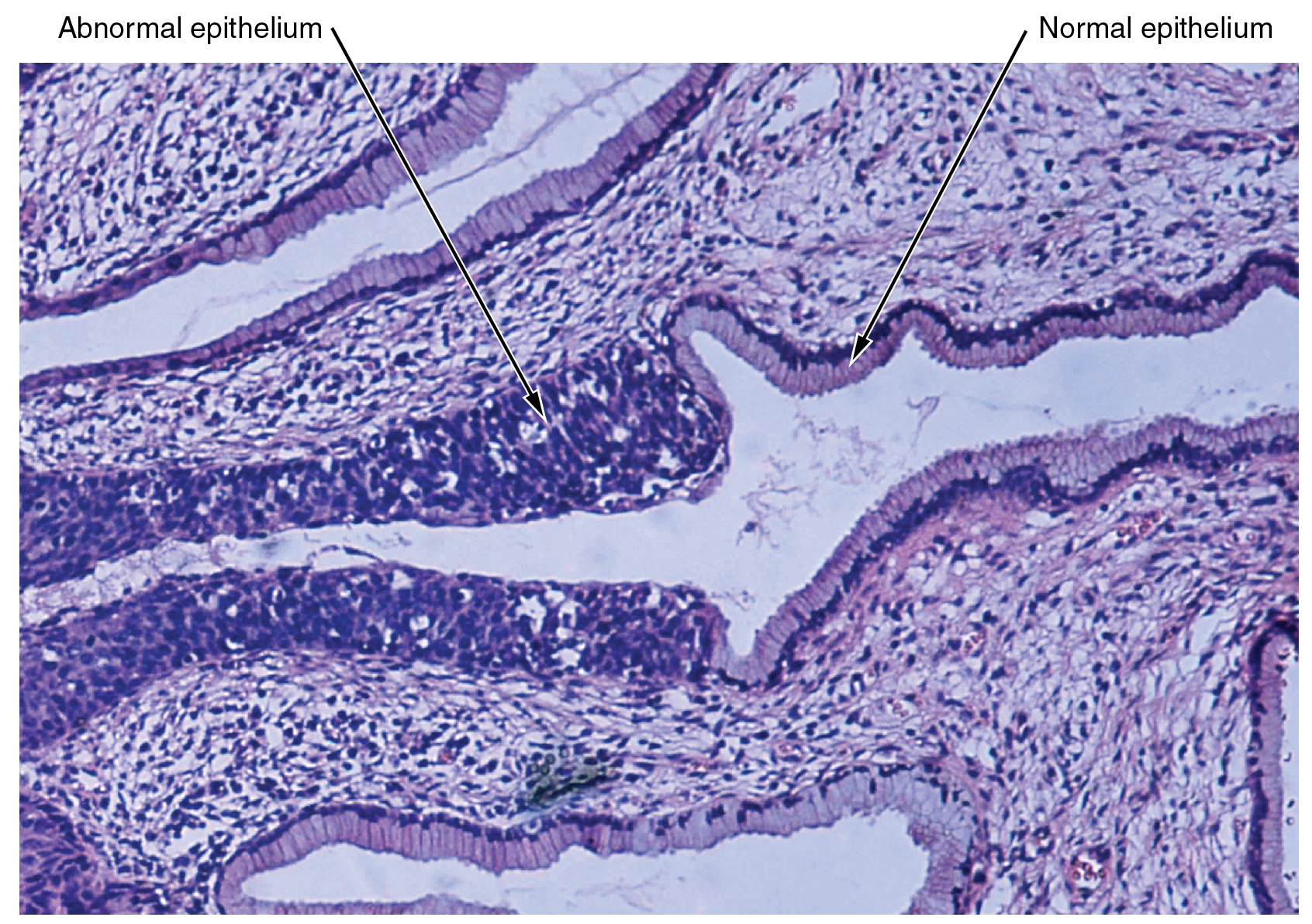
The body contains at least 200 distinct cell types. These cells contain essentially the same internal structures yet they vary enormously in shape and function. The different types of cells are not randomly distributed throughout the body; rather they occur in organized layers, a level of organization referred to as tissue. The micrograph that opens this chapter shows the high degree of organization among different types of cells in the tissue of the cervix. You can also see how that organization breaks down when cancer takes over the regular mitotic functioning of a cell.
The variety in shape reflects the many different roles that cells fulfill in your body. The human body starts as a single cell at fertilization. As this fertilized egg divides, it gives rise to trillions of cells, each built from the same blueprint, but organizing into tissues and becoming irreversibly committed to a developmental pathway.
Question: What is the function of synovial membranes?
Choices:
Synovial membranes are a type of connective tissue membrane that supports mobility in joints. The membrane lines the joint cavity and contains fibroblasts that produce hyaluronan, which leads to the production of synovial fluid, a natural lubricant that enables the bones of a joint to move freely against one another.
Question: Watch this video (http://openstaxcollege.org/l/healinghand) to see a hand heal. Over what period of time do you think these images were taken?
Choices:
Approximately one month.
Question: Identify the four types of tissue in the body, and describe the major functions of each tissue.
Choices:
The four types of tissue in the body are epithelial, connective, muscle, and nervous. Epithelial tissue is made of layers of cells that cover the surfaces of the body that come into contact with the exterior world, line internal cavities, and form glands. Connective tissue binds the cells and organs of the body together and performs many functions, especially in the protection, support, and integration of the body. Muscle tissue, which responds to stimulation and contracts to provide movement, is divided into three major types: skeletal (voluntary) muscles, smooth muscles, and the cardiac muscle in the heart. Nervous tissue allows the body to receive signals and transmit information as electric impulses from one region of the body to another.
Question: View this slideshow (http://openstaxcollege.org/l/stemcells) to learn more about stem cells. How do somatic stem cells differ from embryonic stem cells?
Choices:
Most somatic stem cells give rise to only a few cell types.
Question: Watch this video (http://openstaxcollege.org/l/etissues) to find out more about the anatomy of epithelial tissues. Where in the body would one find non-keratinizing stratified squamous epithelium?
Choices:
The inside of the mouth, esophagus, vaginal canal, and anus.
Question: Follow this link (http://openstaxcollege.org/l/nobel) to learn more about nervous tissue. What are the main parts of a nerve cell?
Choices:
Dendrites, cell body, and the axon.
Question: Watch this video (http://openstaxcollege.org/l/tumor) to learn more about tumors. What is a tumor?
Choices:
A mass of cancer cells that continue to grow and divide.
Question: Watch this video (http://openstaxcollege.org/l/musctissue) to learn more about muscle tissue. In looking through a microscope how could you distinguish skeletal muscle tissue from smooth muscle?
Choices:
Skeletal muscle cells are striated.
Question: The structure of a tissue usually is optimized for its function. Describe how the structure of individual cells and tissue arrangement of the intestine lining matches its main function, to absorb nutrients.
Choices:
Columnar epithelia, which form the lining of the digestive tract, can be either simple or stratified. The cells are long and narrow. The nucleus is elongated and located on the basal side of the cell. Ciliated columnar epithelium is composed of simple columnar epithelial cells that display cilia on their apical surfaces.
Question: The zygote is described as totipotent because it ultimately gives rise to all the cells in your body including the highly specialized cells of your nervous system. Describe this transition, discussing the steps and processes that lead to these specialized cells.
Choices:
The zygote divides into many cells. As these cells become specialized, they lose their ability to differentiate into all tissues. At first they form the three primary germ layers. Following the cells of the ectodermal germ layer, they too become more restricted in what they can form. Ultimately, some of these ectodermal cells become further restricted and differentiate in to nerve cells.
Question: Visit this link (http://openstaxcollege.org/l/10quiz) to test your connective tissue knowledge with this 10-question quiz. Can you name the 10 tissue types shown in the histology slides?
Choices:
Click at the bottom of the quiz for the answers.